Difei Wang1, Rüdiger Stirnberg1, Eberhard Pracht1, and Tony Stöcker1,2
1German Centre for Neurodegenerative Diseases (DZNE), Bonn, Germany, 2Department of Physics and Astronomy, University of Bonn, Bonn, Germany
1German Centre for Neurodegenerative Diseases (DZNE), Bonn, Germany, 2Department of Physics and Astronomy, University of Bonn, Bonn, Germany
Fast MPM is achieved at 7T using 3D-EPI with PTx pulses to reduce B1+
field
inhomogeneities. The weighted PTx images are more homogeneous in the
Cerebellum. In the MPM framework, the residual inhomogeneity can be compensated for by B1+
field correction, resulting in high-quality parameter maps.
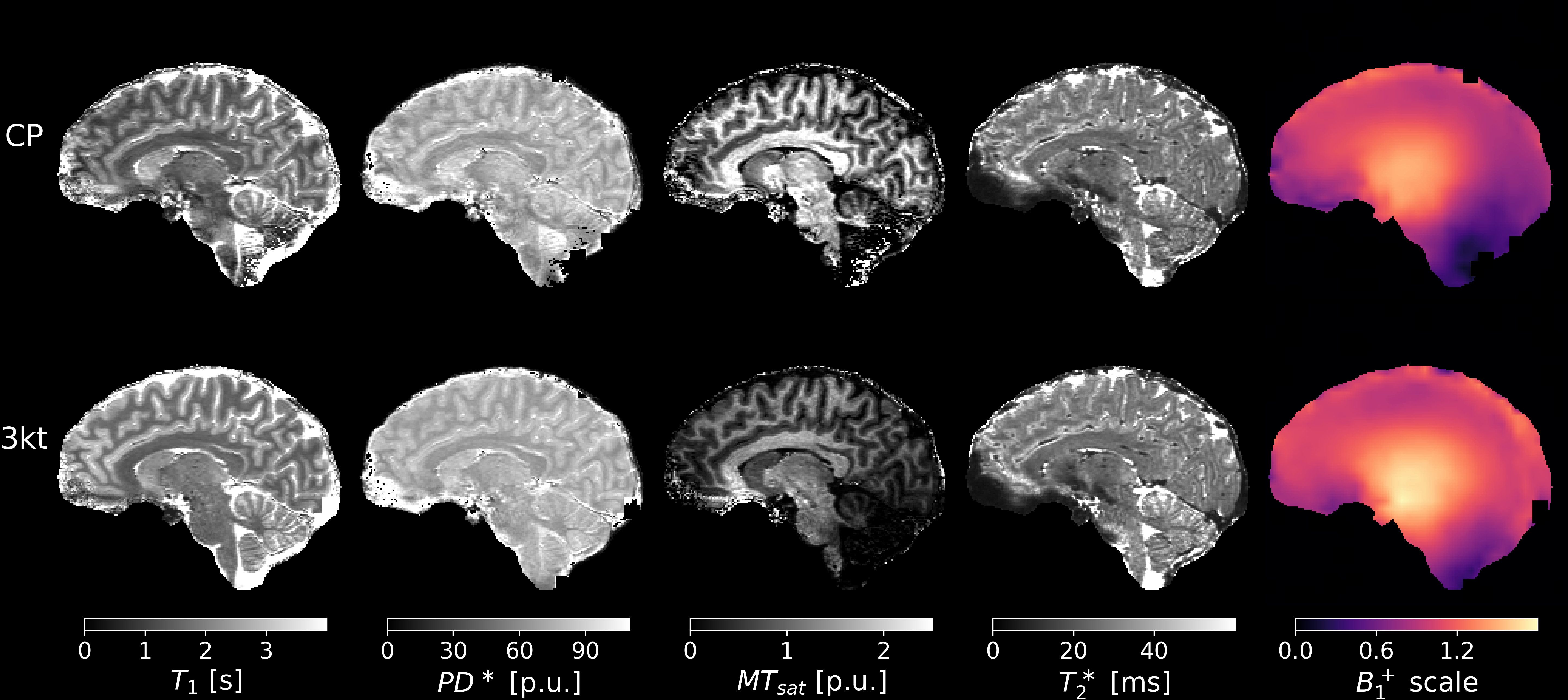
Fig. 3
A sagittal view of T1, PD* , MTsat
and T2* maps using the data acquired with the
CP mode and PTx pulses along with the corresponding B1+
scale map. The TR is 45 ms for both MTw scans. The nominal MT flip
angle is 260° for PTx image and 320° for CP. Both MTsat maps have
low CNR due to the insufficient MT saturation, especially in the
Cerebellum. The PTx B1+ scale map shows improved
excitation in the Cerebellum compared to the CP map. The PTx T1
map has higher values than the CP T1 map and robust voxel
estimates throughout the brain.
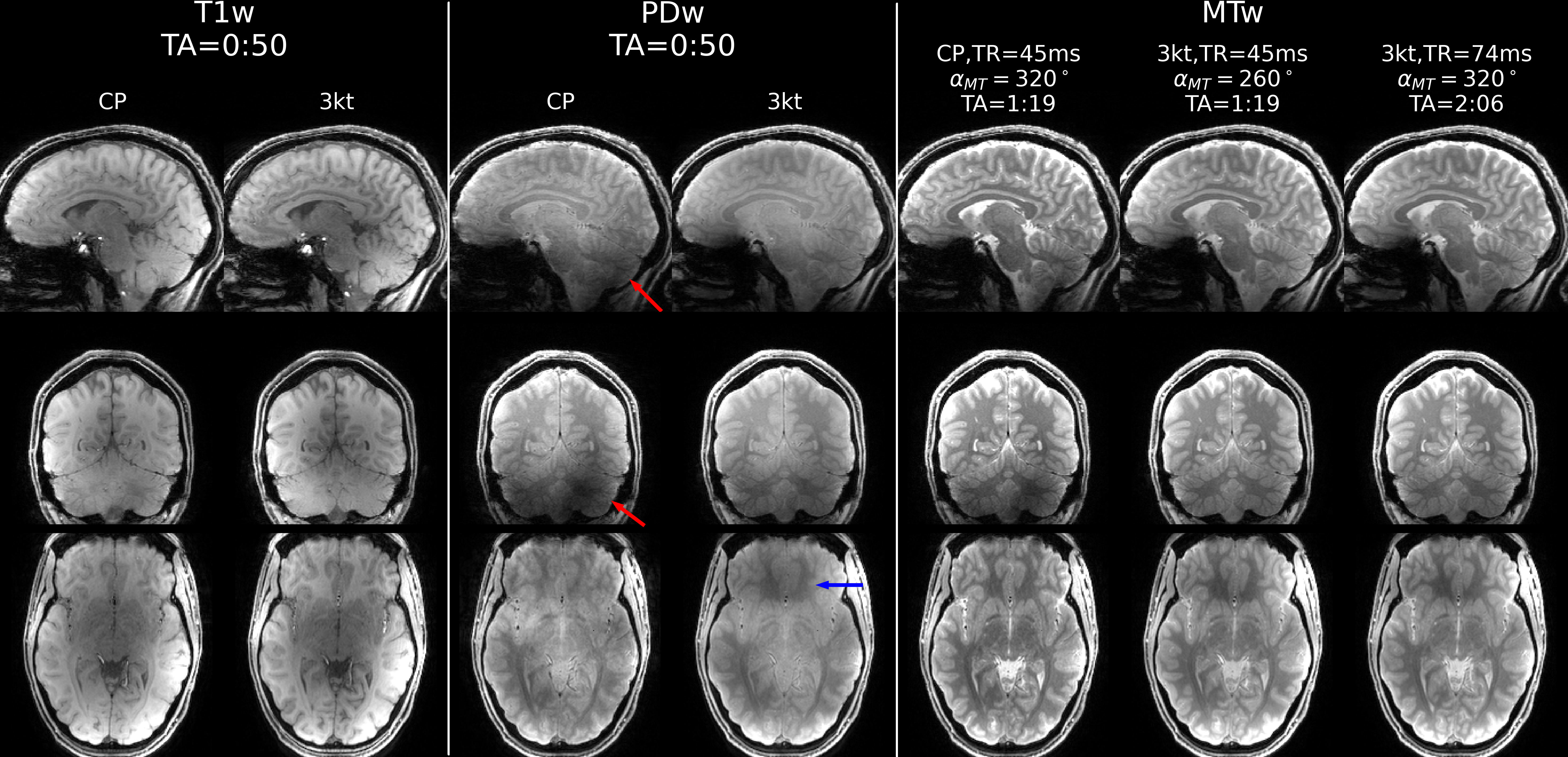
Fig. 1
Sagittal, coronal and axial view of T1w, PDw and MTw
images acquired using CP mode and PTx pulses. The last column shows
the MTw scan with prolonged TR and the same nominal MT flip angle as
the CP mode. The acquisition times are listed for each scan. All
three MTw scans share the same CP mode MT pulse with different
nominal saturation flip angles and TRs in order to match the SAR limits, listed respectively. The ones
with a higher FA show slightly better soft tissue contrast.