Georg Schramm1, Johan Nuyts1, and Fernando Boada2
1KU Leuven, Leuven, Belgium, 2New York University School of Medicine, New York City, NY, United States
1KU Leuven, Leuven, Belgium, 2New York University School of Medicine, New York City, NY, United States
Anatomical regularization and T2* signal decay modeling during readout allow to suppress noise while preserving anatomical detail in reconstructions of dual echo sodium MR data.
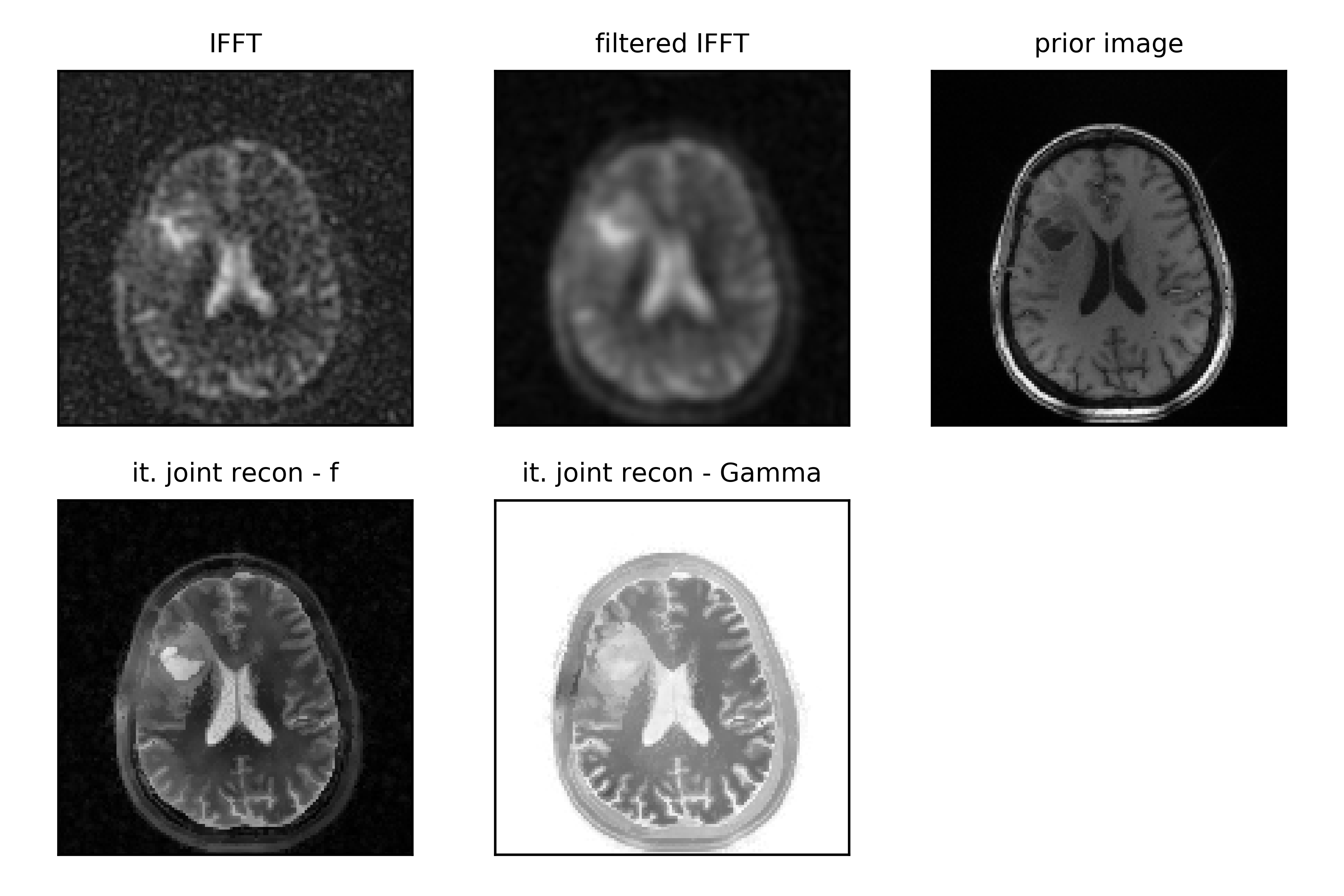
Figure 3: Trans-axial slice of a reconstruction of a real brain tumor data set. (top left) IFFT of data from 1 st echo. (top middle) Han filtered IFFT. (top right) T1 1H image used as anatomical prior. (bottom left) iterative reconstruction of Na signal using anatomical Bowsher prior and signal from 2 echos (βf = 0.01). (bottom middle) jointly reconstructed decay image Γ (βΓ = 0.03).
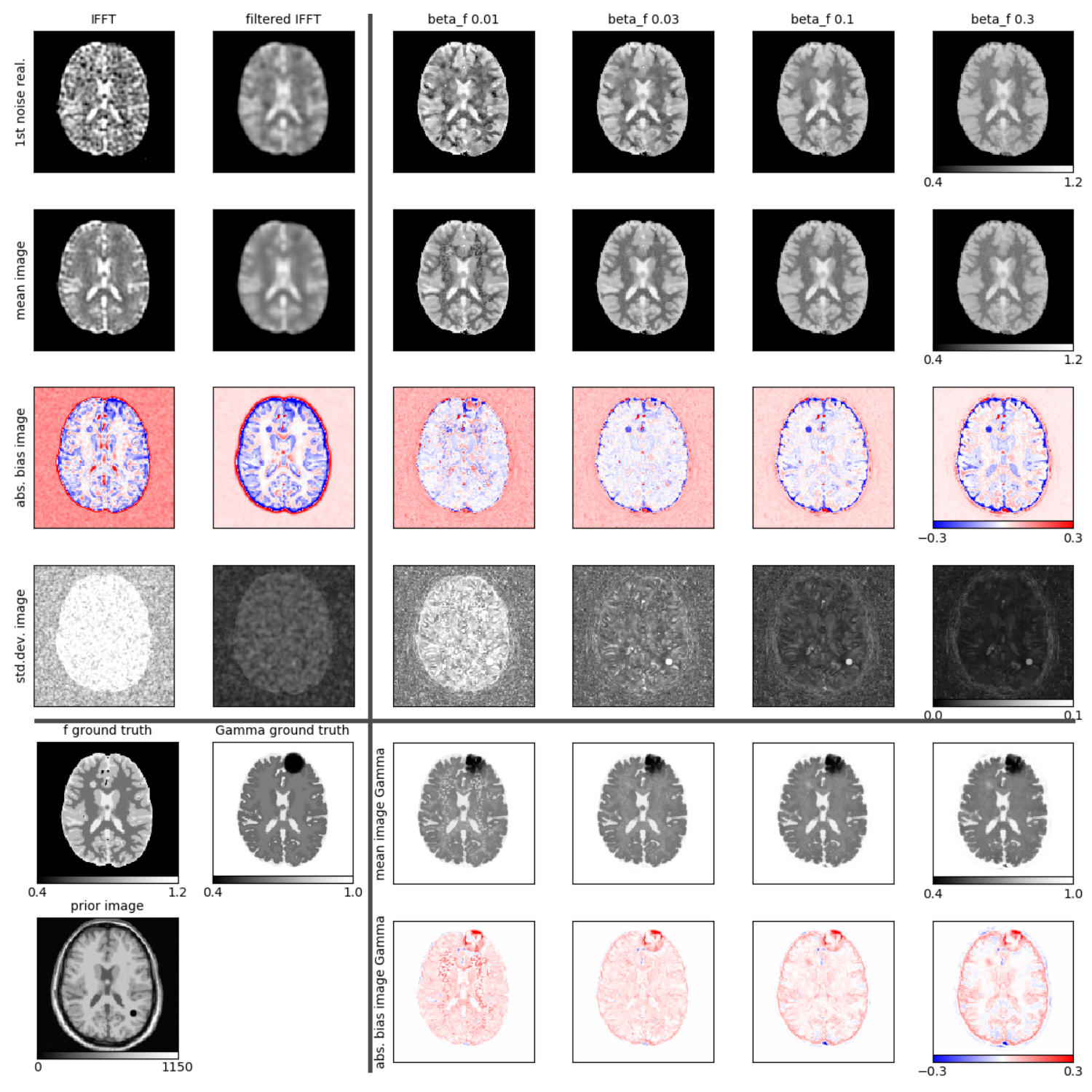
Figure 1: Results of 3D reconstructions of simulated dual echo Na MR data. Reconstruction of first noise realization, mean, bias and standard deviations imagesof f and Γ. (top left) unfiltered and filtered IFFT of data. (bottom left) simulated ground truth images. (right) Reconstructions of f and Γ for different levels of regularization.