Giorgia Milotta1, Nadège Corbin1,2, Christian Lambert1, Antoine Lutti3, Siawoosh Mohammadi4,5, and Martina Callaghan1
1Wellcome Centre for Human Neuroimaging, UCL Queen Square Institute of Neurology, University College London, London, United Kingdom, 2Centre de Résonance Magnétique des Systèmes Biologiques, UMR5536, CNRS/University Bordeaux, Bordeaux, France, 3Laboratory for Research in Neuroimaging, Department for Clinical Neuroscience, Lausanne University Hospital and University of Lausanne, Lausanne, Switzerland, 4Department of Systems Neurosciences, University Medical Center Hamburg-Eppendorf, Hamburg, Germany, 5Department of Neurophysics, Max Planck Institute for Human Cognitive and Brain Sciences, Leipzig, Germany
1Wellcome Centre for Human Neuroimaging, UCL Queen Square Institute of Neurology, University College London, London, United Kingdom, 2Centre de Résonance Magnétique des Systèmes Biologiques, UMR5536, CNRS/University Bordeaux, Bordeaux, France, 3Laboratory for Research in Neuroimaging, Department for Clinical Neuroscience, Lausanne University Hospital and University of Lausanne, Lausanne, Switzerland, 4Department of Systems Neurosciences, University Medical Center Hamburg-Eppendorf, Hamburg, Germany, 5Department of Neurophysics, Max Planck Institute for Human Cognitive and Brain Sciences, Leipzig, Germany
With a
heuristic linear model of the FA-dependence of R2* we observed
excellent agreement between empirical observations and simulations based on two
exchanging water pools. Results indicate that the FA-dependence is greatest for
high myelin water fraction and slow exchange.
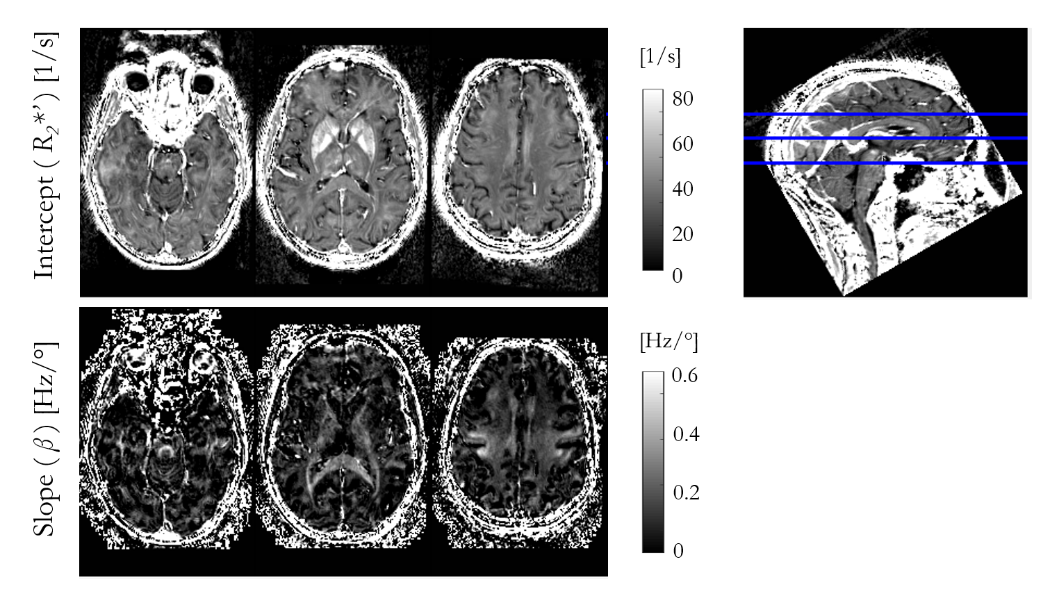
Figure 2 – Intercept (β) and
slope (R2*’) of R2* FA dependence obtained from linear
fitting according to Eq. 1. The same axial slices are shown as in Figure 1. The
spatial variability in the slope (β) highlights
specific, predominantly white matter structures, such as the superior
cerebellar fibres, brachium conjunctivum and corpus callosum.
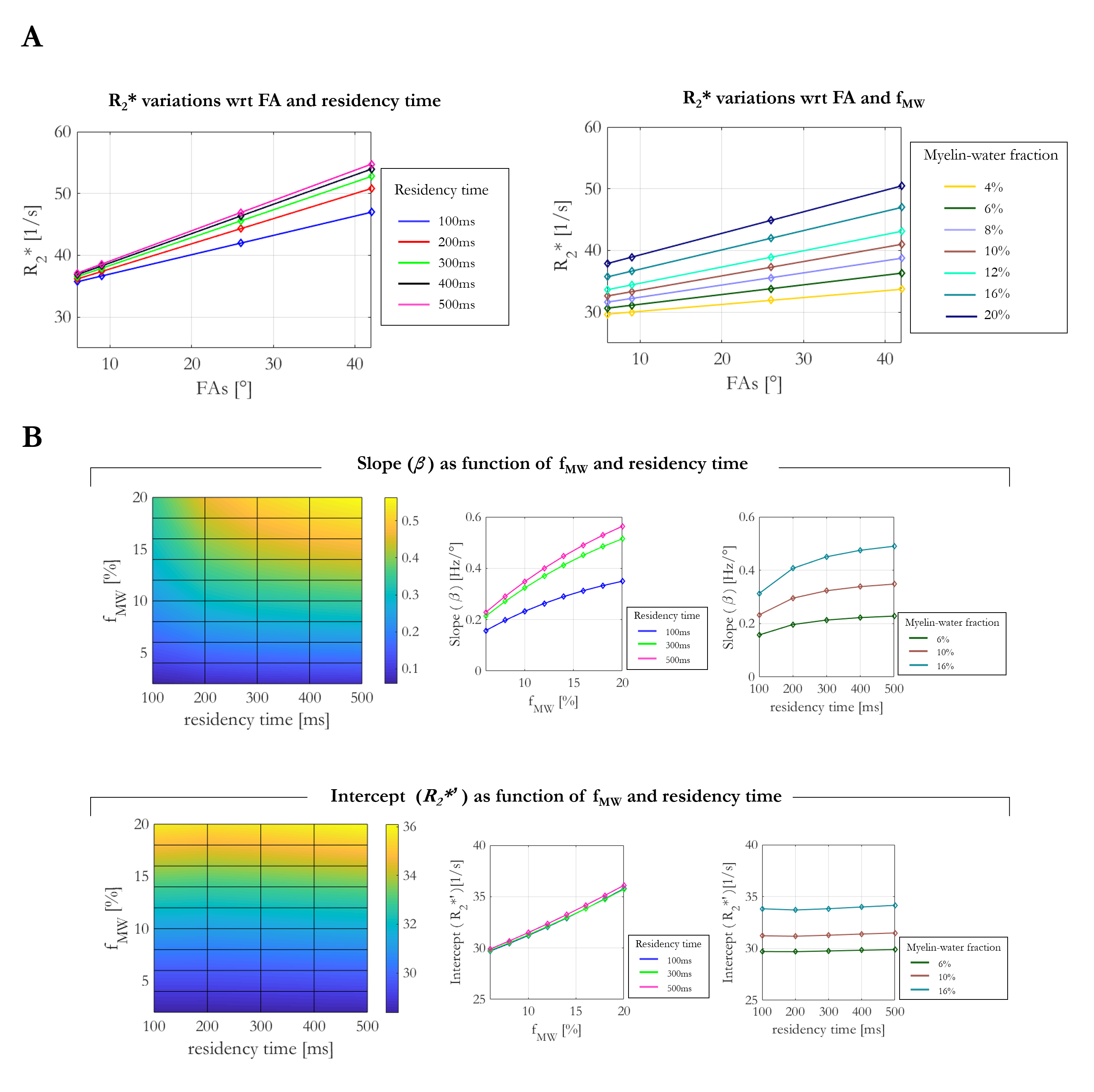
Figure 3 – Simulations of R2* variation with respect to FA,
residency time and fMW. A: R2* as a function of FA
together with linear fits according to Eq. 1 for a fixed fMW of 16%,
or a fixed residency time of 100ms. B: Variability of slopes and intercepts across
investigated conditions (residency time = 100-500ms, fMW = 2-20%).
The intercept R2*’ shows higher sensitivity to fMW
(12.6%), but effectively no dependence on residency time (0.74% variance across
range investigated). The slope was most sensitive to the fMW (~55%),
but additionally depended on residency time (~13%).