Gunther Helms1 and Hampus Olsson1
1Medical Radiation Physics, Clinical Sciences Lund, Lund University, Lund, Sweden
1Medical Radiation Physics, Clinical Sciences Lund, Lund University, Lund, Sweden
Preparing
a driven equilibrium prior to inversion by an MP2RAGE sequence allows for IR-based
T1 mapping of the whole brain, thus providing a B1+-independent, T1
reference for quantitative studies.
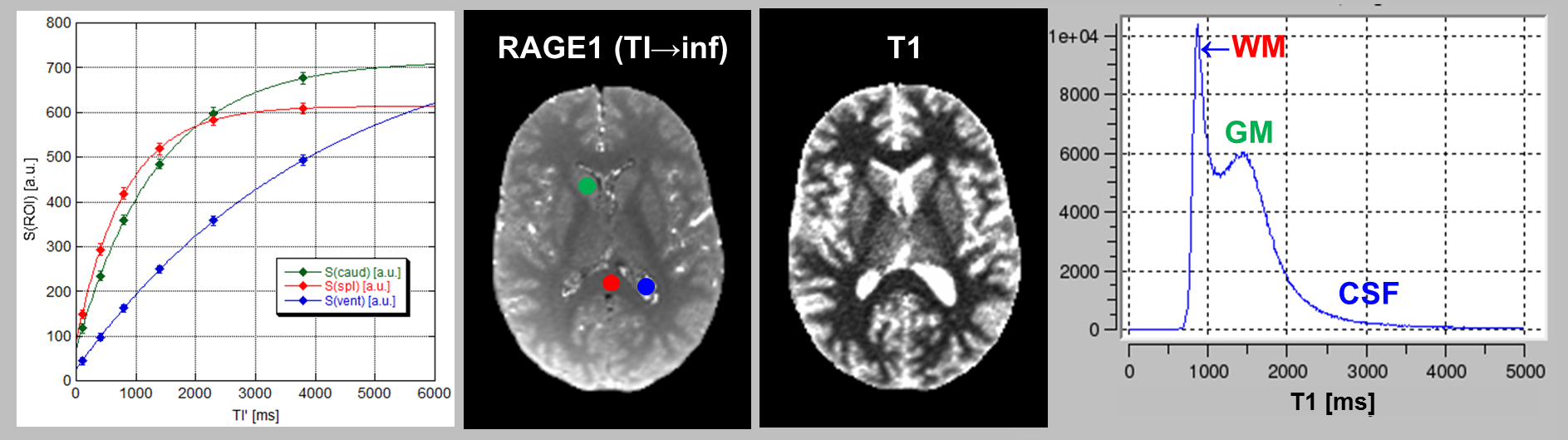
Figure
2: Left: Exponential fits to ROIs in caudate (green),
splenium (red) and lateral ventricle (blue). S(0) represents the
intensity of the DE after inversion and partial convergence at readout of k-space center. Center: Maps of
extrapolated RAGE intensity and T1 after coregistration of RAGE2 volumes.
Right: Whole brain histogram of fitted T1 values at 3T.
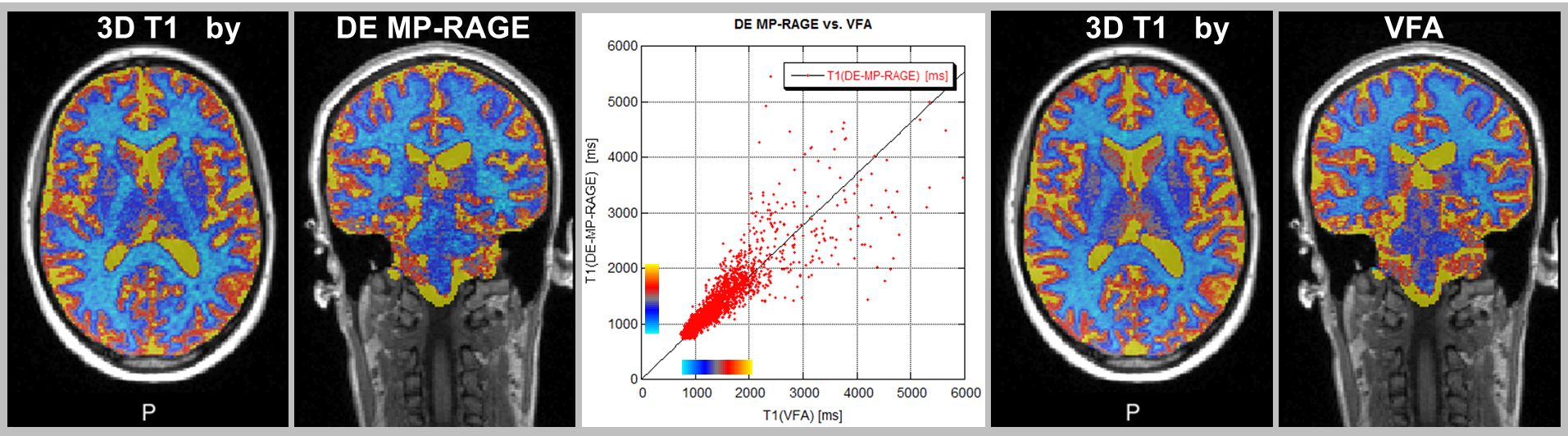
Figure
3: Comparison
of T1 obtained by DE MP-RAGE (left) with those obtained by VFA at constant
B1=8.16uT (right). Scatterplot of 2800 pixels revealed slightly lower T1 by VFA
(0.925±0.005). The pseudo-color overlay indicates slightly larger noise in the unbiased DE
MP-RAGE maps.