Gian Franco Piredda1,2,3, Tom Hilbert1,2,3, Berkin Bilgic4,5,6, Erick Jorge Canales-Rodríguez3, Marco Pizzolato3,7, Reto Meuli2, Jean-Philippe Thiran2,3, and Tobias Kober1,2,3
1Advanced Clinical Imaging Technology, Siemens Healthcare AG, Lausanne, Switzerland, 2Department of Radiology, Lausanne University Hospital and University of Lausanne, Lausanne, Switzerland, 3LTS5, École Polytechnique Fédérale de Lausanne (EPFL), Lausanne, Switzerland, 4Athinoula A. Martinos Center for Biomedical Imaging, Charlestown, MA, United States, 5Department of Radiology, Harvard Medical School, Boston, MA, United States, 6Harvard‐MIT Health Sciences and Technology, MIT, Cambridge, MA, United States, 7Department of Applied Mathematics and Computer Science, Technical University of Denmark, Kongens Lyngby, Denmark
1Advanced Clinical Imaging Technology, Siemens Healthcare AG, Lausanne, Switzerland, 2Department of Radiology, Lausanne University Hospital and University of Lausanne, Lausanne, Switzerland, 3LTS5, École Polytechnique Fédérale de Lausanne (EPFL), Lausanne, Switzerland, 4Athinoula A. Martinos Center for Biomedical Imaging, Charlestown, MA, United States, 5Department of Radiology, Harvard Medical School, Boston, MA, United States, 6Harvard‐MIT Health Sciences and Technology, MIT, Cambridge, MA, United States, 7Department of Applied Mathematics and Computer Science, Technical University of Denmark, Kongens Lyngby, Denmark
The proposed joint-CAIPI reconstruction across echoes of GRASE data delivers myelin water fraction maps with mitigated aliasing artifacts and ~40% reduction in scan time in comparison to prior CAIPIRINHA acquisition and reconstruction strategies.

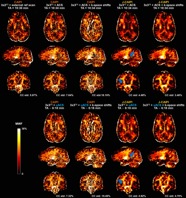
Figure 3. Example MWF maps obtained from T2-weighted contrasts acquired with different k-space sampling schemes or with retrospective undersampling of the ACS region (uACS) and reconstructed with conventional and joint-CAIPI (J-CAIPI). Residual aliasing artifacts in CAIPI reconstructions with integrated ACS heavily compromise image quality of retrieved MWF maps. Such artifacts are mitigated in J-CAIPI reconstructions but still present if k-space shifts across echoes are not employed (blue arrows). MWF std in corpus callosum (CC) decreases in J-CAIPI reconstructions.

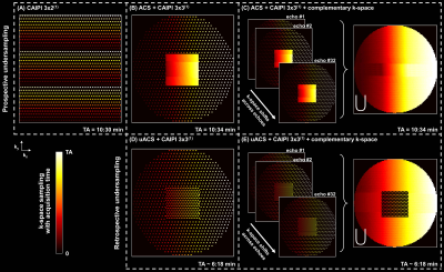
Figure 1. k-space sampling strategies investigated in this study. (A) CAIPI 3x2(1) (acquired with an external reference scan for calibrating the reconstruction kernels). (B) CAIPI 3x3(1) on an elliptical Cartesian grid with an integrated autocalibration signal (ACS) region and (C) with additional shifts across echoes for complementary k-space coverage. (D, E) Same as in (B) and (C), respectively, but with retrospective undersampling of the ACS region with a CAIPI 2x2(1) pattern. Acquisition times (TAs) are given for a 1.6 mm3 isotropic whole-brain scan.
