Fardad Michael Serry1, Junzhou Chen1,2, Anthony G Christodoulou1,2, Yuheng Huang1,2, Fei Han3, Won Bae4,5, Christine Chung4,5, Richard Handlin1, John Stager1, Matthew Dausch1, Yubin Cai1, Yujie Shan1, Yucen Liu1, Yibin Xie1, Xiaoming Bi3, Rohan Dharmakumar1,2, Zhaoyang Fan1,6, Debiao Li1,2, Hsin-Jung Yang1, and Hui Han1
1Biomedical Imaging Research Institute, Cedars-Sinai Medical Center, Los Angeles, CA, United States, 2Department of Bioengineering, UCLA, Los Angeles, CA, United States, 3Siemens Medical Solutions USA, Inc., Los Angeles, CA, United States, 4University of California, San Diego, San Diego, CA, United States, 5VA Medical Center, San Diego, CA, United States, 6University of Southern California Department of Radiology, Los Angeles, CA, United States
1Biomedical Imaging Research Institute, Cedars-Sinai Medical Center, Los Angeles, CA, United States, 2Department of Bioengineering, UCLA, Los Angeles, CA, United States, 3Siemens Medical Solutions USA, Inc., Los Angeles, CA, United States, 4University of California, San Diego, San Diego, CA, United States, 5VA Medical Center, San Diego, CA, United States, 6University of Southern California Department of Radiology, Los Angeles, CA, United States
Novel unified
coil (UNIC) array 3D B0 shim reduced metal-induced signal void artifact,
increasing the visible area around a hip prosthesis in phantom by up to
50% in some slices. The sequence-agnostic technology was tested with a 3D GRE, generally more susceptible to metal artifact than SE.

Figure 3. Phantom axial GRE images of cross sections at the femoral head (a,b) and near the femoral insertion end (c,d) of the metal hip prosthesis at TE=4.80ms before (left) and after (right) adding UNIC shim to scanner shim. Signal void is stronger at the longer echo time due to additional dephasing between the two echos. UNIC shimming reduced signal void artifact area by 9% (b versus a) and 9% (d versus c), and extended the visible area closer to the metal, increasing it by 50% (b versus a); and 8 % (d versus c); see text for ROI definition. Fiducial markers are visible in a and b.
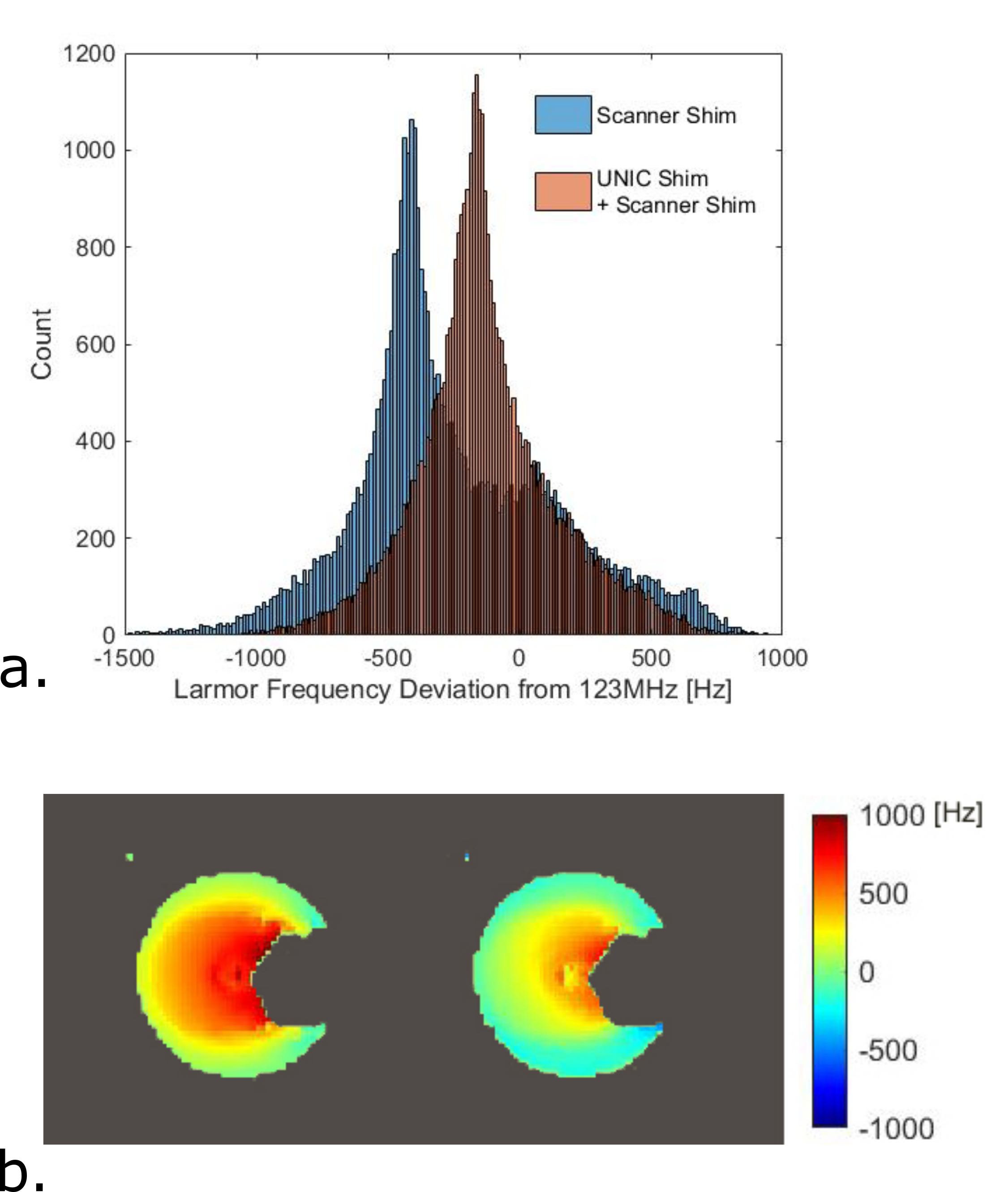
Figure 5. B0 field homogeneity improvement
with UNIC shimming. Histograms of Larmor frequency deviation from the scanner center
frequency (~123MHz), of the masked volume of pixels in the 3D slab scanned before
and after adding UNIC shim to scanner shim (a), and B0 field magnitude maps (b)
for the slice in Figures 3c,d before (b. left) and after (b. right) adding UNIC
shim to scanner shim.