Matthew A. McCready1, William B. Handler1, Francisco Martinez2,3, Timothy J. Scholl2,3, and Blaine A. Chronik1,2
1Physics and Astronomy, Western University, London, ON, Canada, 2Medical Biophysics, Western University, London, ON, Canada, 3Robarts Research Institute, Western University, London, ON, Canada
1Physics and Astronomy, Western University, London, ON, Canada, 2Medical Biophysics, Western University, London, ON, Canada, 3Robarts Research Institute, Western University, London, ON, Canada
The dreMR method assumes field strength is homogenous and coils reach field instantly. We have shown that these assumptions result in errors within dreMR images, which can be solved by use of improved homogeneity coils and higher slew rates.
Figure
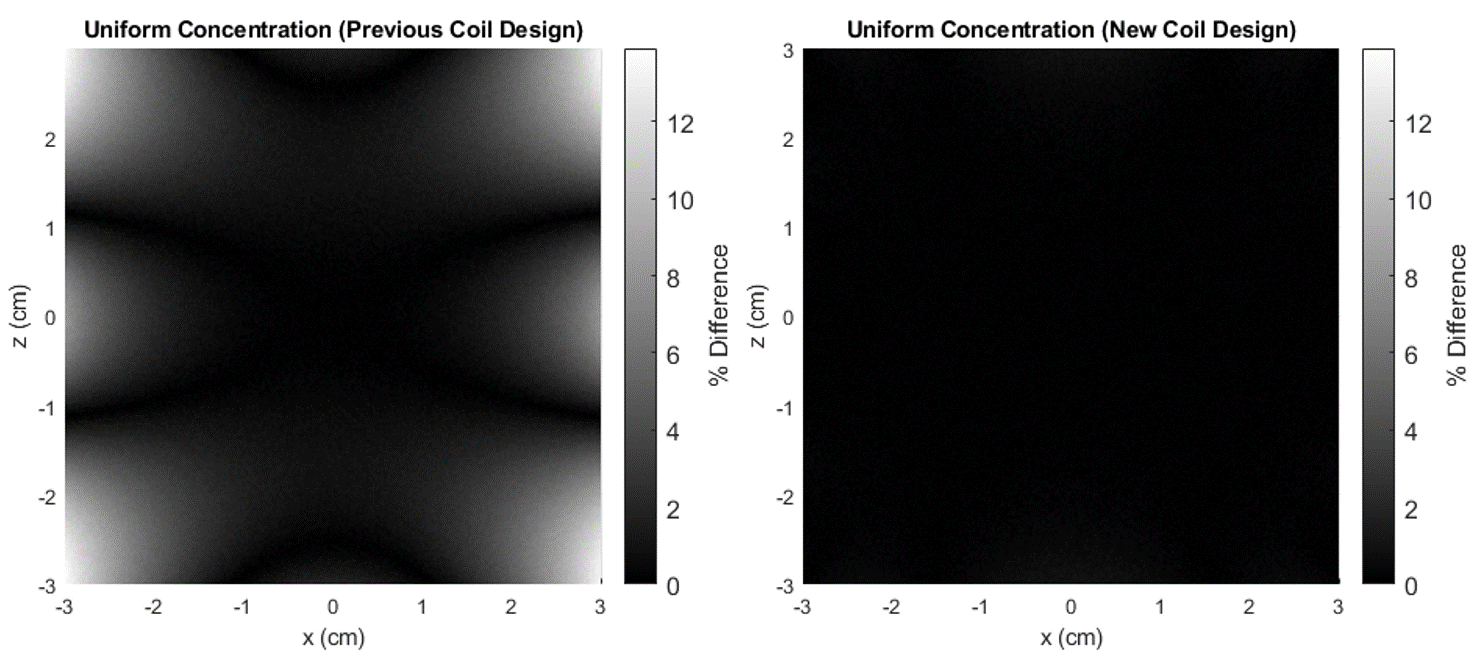
2. (LEFT) Percent difference in dreMR image of VivoTrax, in uniform
concentration of 160 μM, using field of previously constructed coil. Percent
difference is taken from center point where field is at desired value. (RIGHT) Same
simulation using field from new design method coil. Colour axis chosen to match
the left.
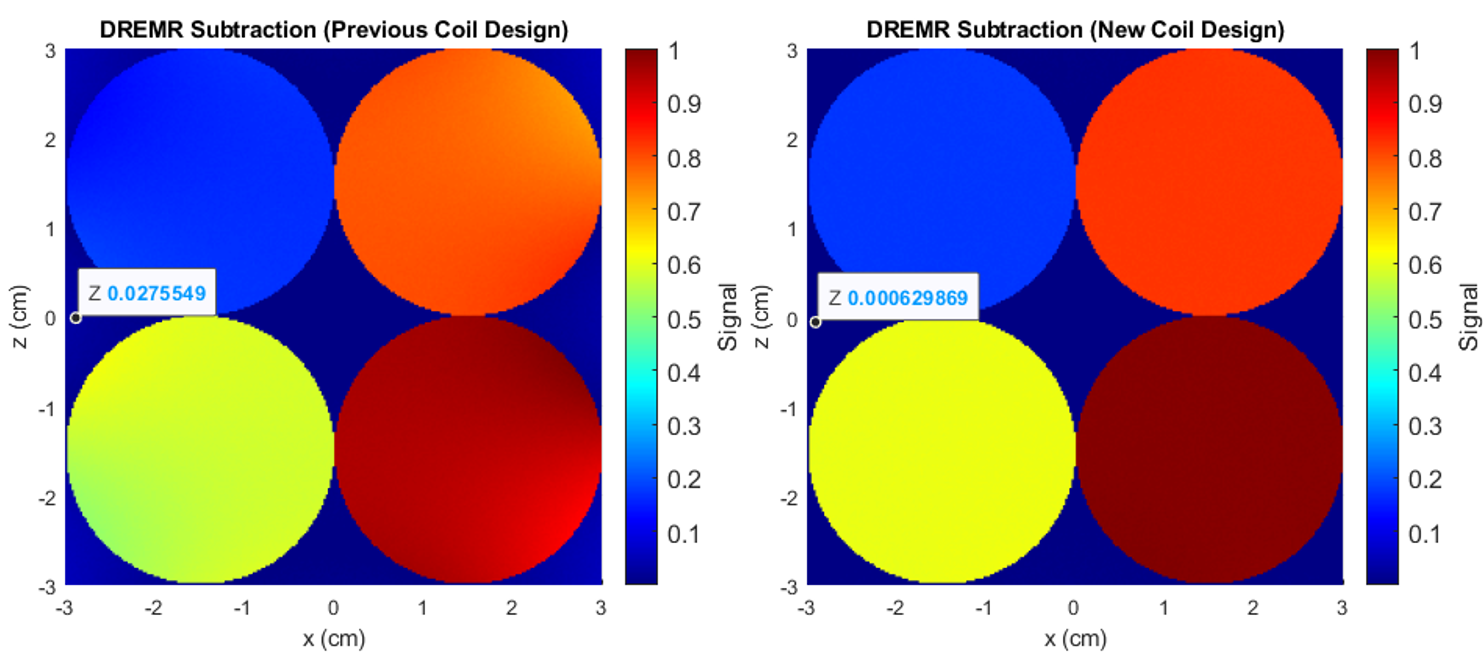
Figure 1. (LEFT) Simulated dreMR image of 4 cylinders of VivoTrax, in concentrations of 20, 80, 120, and 160 μM, using field of previously constructed coil. Note change in signal across cylinders. Sequence uses 300mT field shift in a 0.5T system. (RIGHT) Same simulation using field from new design method coil. Signal does not noticeably change across cylinders. Data points are given in both figures at locations with no contrast agent.