Robert Kowal1, Enrico Pannicke2, Marcus Prier1, Ralf Vick2, Georg Rose3, and Oliver Speck1
1Department of Biomedical Magnetic Resonance, Otto von Guericke University, Magdeburg, Germany, 2Chair of Electromagnetic Compatibility, Otto von Guericke University, Magdeburg, Germany, 3Chair in Healthcare Telematics and Medical Engineering, Otto von Guericke University, Magdeburg, Germany
1Department of Biomedical Magnetic Resonance, Otto von Guericke University, Magdeburg, Germany, 2Chair of Electromagnetic Compatibility, Otto von Guericke University, Magdeburg, Germany, 3Chair in Healthcare Telematics and Medical Engineering, Otto von Guericke University, Magdeburg, Germany
A low-field (0.26T) Tabletop-MR-system was compared to a high-field (3T) clinical scanner by evaluating the SNR of simple FID time domain signals. Direct comparison of the SNR led to a measured relative performance of 11.3% for the Tabletop before compensation for experimental conditions.
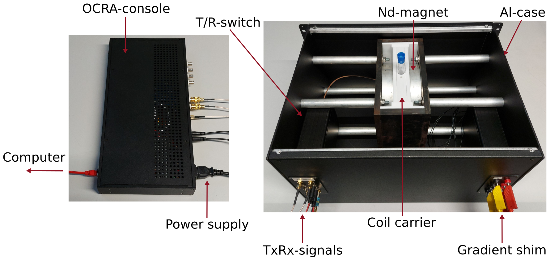
Measurement setup on the Tabletop-system. Left side: OCRA-console for controlling the system, supplying the components and connecting to the computer. Right side: Opened system with the sample in the carrier between the neodymium magnets with connected signal and shimming lines.
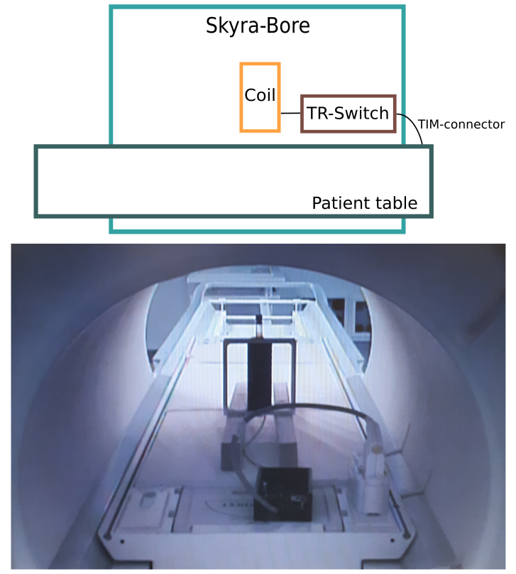
Measurement setup on the Skyra-system. The test tube sample was positioned with the coil perpendicular to the static magnetic
field
fixed by wooden planks on the patient table. The connection to the MR system was established via the T/R-switch and TIM-connector. Shown on the bottom is an image taken with the patient camera.