Zhenhua Shen1, Xuchen Zhu1, Shihong Han1, Fuyi Fang1, Wei Luo1, Shao Che1, Zidong Wei1, Jinguang Zong1, Yongquan Ye2, Bo Li1, Shuheng Zhang1, Anthony Vu2, Weiguo Zhang2, and Guobin Li1
1United Imaging Healthcare, Shanghai, China, 2UIH America, Inc., Houston, TX, United States
1United Imaging Healthcare, Shanghai, China, 2UIH America, Inc., Houston, TX, United States
With
multi-channel RF parallel transmission hardware architecture and static RF shimming
techniques, the uniformity of the RF transmission field is shown to be well
controlled for imaging quality guarantee. Preliminary results show great promise
for body imaging at 5T.
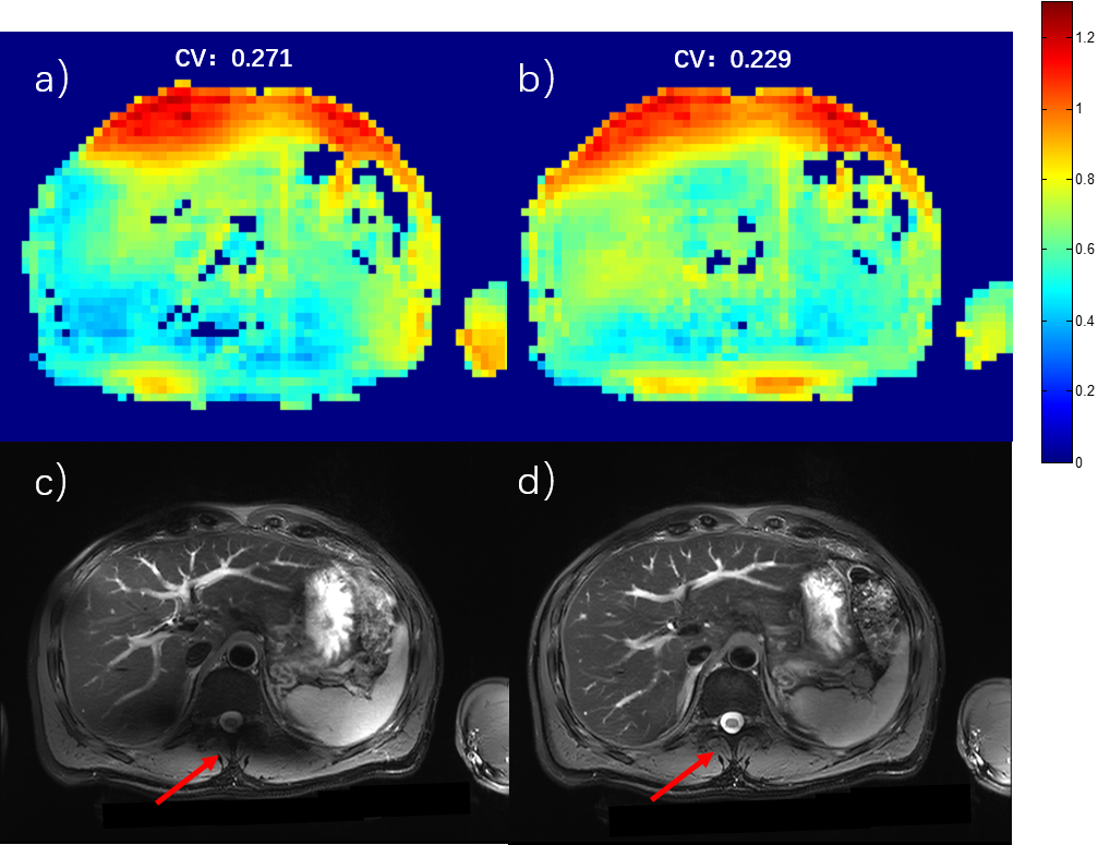
Figure 2 Abdominal imaging at 5T. a) and c) B1+ sensitivity map and T2W FSE image with CP mode excitation. b) and d) B1+sensitivity map and T2W FSE image after static RF shimming optimization. The CV values were measured for bothcases.The red arrows show the most significant improvement area.
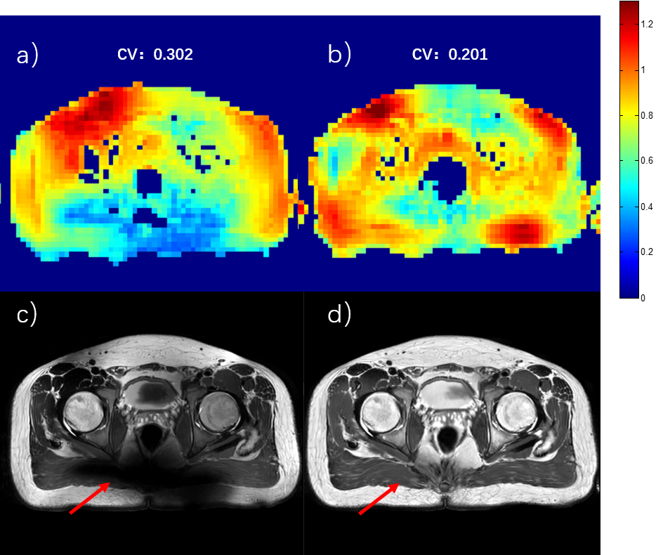
Figure 3 Pelvic imaging at 5T. a) and c) B1+ sensitivity map and T2W FSE image with CP mode excitation. b) and d) B1+sensitivity map and T2W FSE image after static RF shimming optimization. The CV values were measured for bothcases.The red arrows show the most significant improvement area.