Sebastian Walter Rieger1, Karla Miller1, Peter Jezzard1, and Wenchuan Wu1
1Wellcome Centre for Integrative Neuroimaging, University of Oxford, Oxford, United Kingdom
1Wellcome Centre for Integrative Neuroimaging, University of Oxford, Oxford, United Kingdom
In
post mortem MRI, RF absorption can cause significant heating in the sample. This
can interfere with scanning and accelerate decomposition of unfixed samples. Here,
a temperature control system is presented which enables prolonged scanning at a
stable temperature while preserving tissue.

Cooling
system schematic. A: Pre-cooling / setup configuration - The chiller is
operating and pre-cooling the bulk of the fluid to the desired temperature,
while the cooling pad and temperature sensor(s) are being used for sample
preparation. B: Normal operation configuration - The temperature probe(s)
are affixed to the sample, and the cooling pad is wrapped around it. A layer of
cloth around the outside provides thermal insulation to prevent excessive
condensation, and the pad is connected to the chiller.
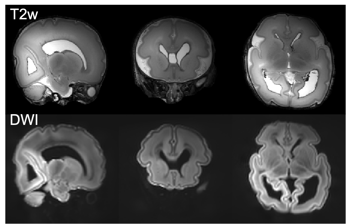
Example T2- and diffusion
weighted images. Top: T2 weighted images were acquired
at 0.4mm isotropic resolution using a 3D-SPACE sequence with FOV=150x150x96mm3,
TR/TE=2300/586ms, 6 repetitions. Bottom: averaged diffusion weighting images
acquired at 0.8mm isotropic resolution using a spin-echo diffusion weighted
readout-segmented EPI sequence with FOV=150x150x104mm3,
TR/TE=10.8s/113ms, multiband factor 2, GRAPPA factor 2, b=9000 s/mm2.