Bruno Sa de La Rocque Guimaraes1, Khaled Talaat1, and Stefan Posse2,3
1Nuclear Engineering, U New Mexico, Albuquerque, NM, United States, 2Neurology, U New Mexico, Albuquerque, NM, United States, 3Physics and Astronomy, U New Mexico, Albuquerque, NM, United States
1Nuclear Engineering, U New Mexico, Albuquerque, NM, United States, 2Neurology, U New Mexico, Albuquerque, NM, United States, 3Physics and Astronomy, U New Mexico, Albuquerque, NM, United States
A novel spectrally and temporally segmented regression approach
for high-speed resting-state fMRI data substantially reduced physiological noise,
motion effects and artificial high-frequency correlations compared with a recently
developed sliding window regression approach.
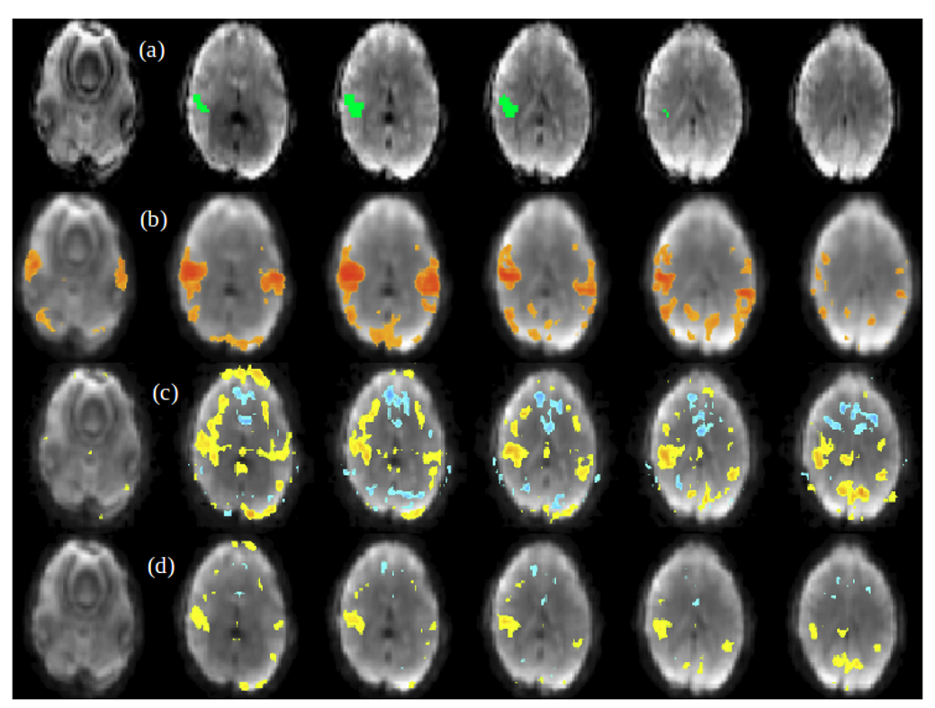
Figure 4. (a) Unilateral AUD seed location in healthy
subject 2, (b) low-frequency resting-state connectivity map thresholded at 0.6,
(c) HRAN regressed data high-frequency resting-state connectivity maps and (d)
spectrally and temporally segmented regressed high-frequency resting-state
connectivity map, both thresholded at 0.3.
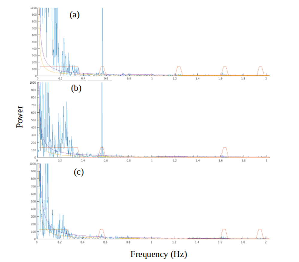
Figure
5. Frequency power spectrum using visual seed in healthy subject 1 visualized
using Turbofilt. (a) Shows the spectrum before regression, (b) shows the
spectra after using HRAN and (c) is the spectrum after performing the spectrally
and temporally segmented regression. The boxes correspond to labels of
physiological noise. The curves correspond to tentative fits of the noise
spectral power. The strong peak at 0.566Hz is a machine artifact.