hacer dasgin1, naciye vardar yagli2, melda saglam2, and kader karli oguz3
1National Magnetic Resonance Research Center (UMRAM), Bilkent University, Ankara, Turkey, 2The Faculty of Physical Therapy and Rehabilitation, Hacettepe University, Ankara, Turkey, 3Faculty of Medicine, Department of Radiology, Hacettepe University, Ankara, Turkey
1National Magnetic Resonance Research Center (UMRAM), Bilkent University, Ankara, Turkey, 2The Faculty of Physical Therapy and Rehabilitation, Hacettepe University, Ankara, Turkey, 3Faculty of Medicine, Department of Radiology, Hacettepe University, Ankara, Turkey
After breath training,
the treatment IMT shows less activation clusters with less activation strength
compared to the sham IMT. Our results agree with previous meditation studies including breathing training, showing
that DMN is deactivated and network gets more organised and localised
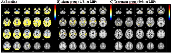
Fig.1. RS-fMRI global correlations of fourteen subjects obtained at baseline
(A) and at 8 weeks after the training at 15% of MIP (B) and profoundly decreased
connectivity patterns at 60% of MIP
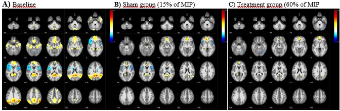
Fig. 2. Compared with Default-mode-network
(DMN) activity at baseline (A), at 8 weeks after breath-training at 15% of MIP
(B) reduced activation (yellow-to-red) at ACC, PCC and left intraparietal
sulcus (IPS) along with diminutive activation at right IPS.
Deactivation (blue) is seen in the bilateral insula and periventricular area
(B). Note that reduction in BOLD activation at ACC network and at posterior
cingulate-retrosplenial cortex, disappeared at the temporal lobes at 60% of MIP
(C)