Helene L Philogene-Khalid1,2, Eric M Cunningham1, Mary F Morrison1,2, and Nicolas R Bolo3,4
1Psychiatry, Lewis Katz School of Medicine at Temple University, Philadelphia, PA, United States, 2Center for Substance Abuse Research, Lewis Katz School of Medicine at Temple University, Philadelphia, PA, United States, 3Psychiatry, Beth Israel Deaconess Medical Center, Boston, MA, United States, 4Psychiatry, Harvard Medical School, Boston, MA, United States
1Psychiatry, Lewis Katz School of Medicine at Temple University, Philadelphia, PA, United States, 2Center for Substance Abuse Research, Lewis Katz School of Medicine at Temple University, Philadelphia, PA, United States, 3Psychiatry, Beth Israel Deaconess Medical Center, Boston, MA, United States, 4Psychiatry, Harvard Medical School, Boston, MA, United States
In cocaine addiction, repeated CLAV decreased the anterior
cingulate connectivity with default mode and cue reactivity related networks,
while it increased the anterior cingulate connectivity with sensory-motor and
motor control processing networks.
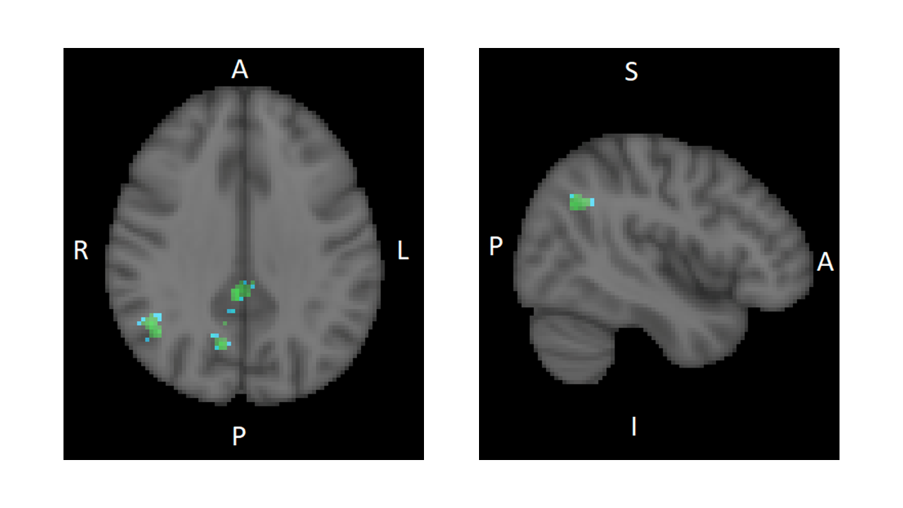
Fig. 1 Statistical nonparametric map showing regions with significant decrease
in functional connectivity (FC) with the ACC after 10days of CLAV
administration compared to baseline. Regions of decreased FC (FDR=0.01,
corrected) in blue-green scale (paired t-test T-value threshold 5.3) overlaid
on the standard MNI-152 brain in grey scale. Right side sagittal slice (y=-56) shows
the angular gyrus region. Left
side axial slice (z=33) shows the angular
gyrus, precuneus, and posterior cingulate cortex regions. R=right
L=left A=anterior P=posterior I=inferior S=superior
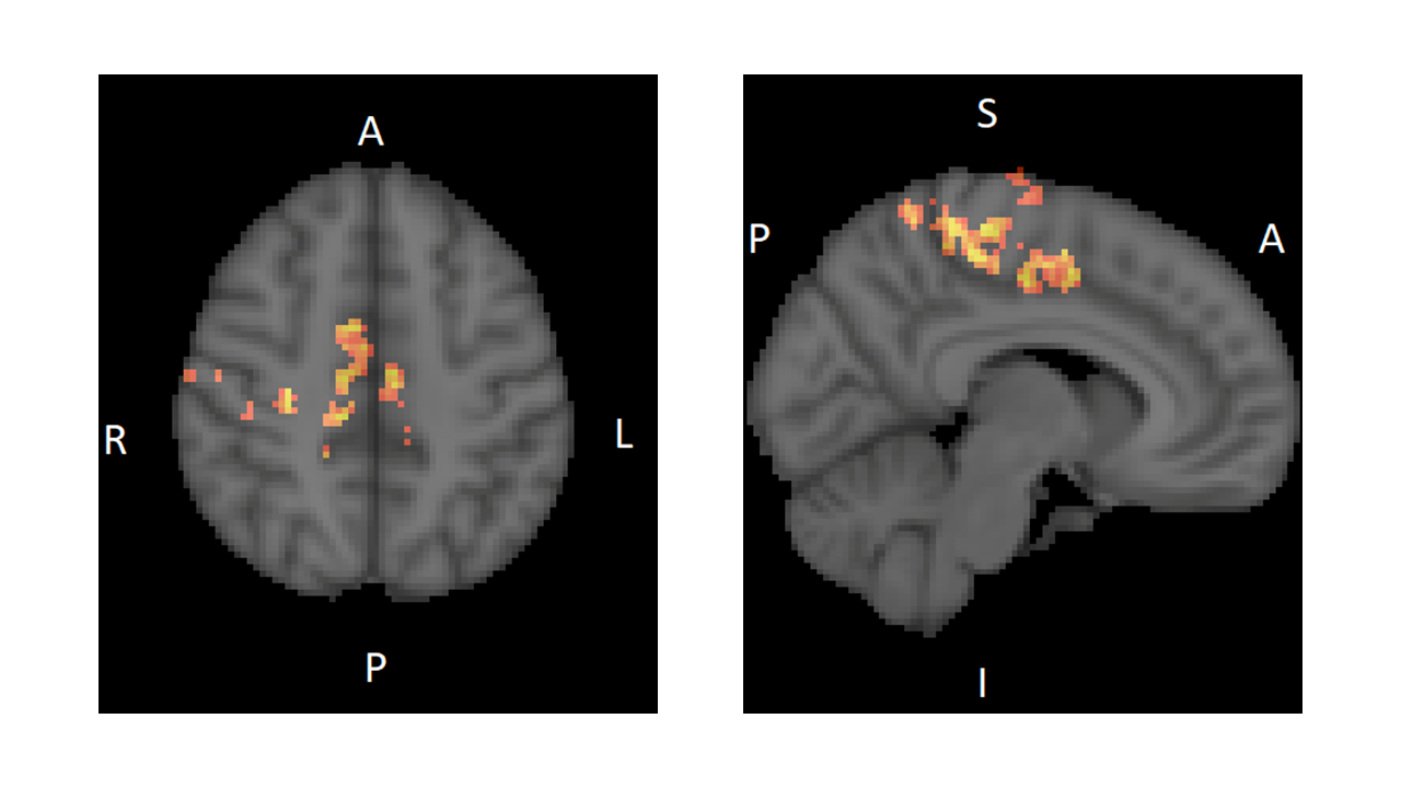
Fig. 2 Statistical nonparametric map showing regions with significant increase
in functional connectivity (FC) with the ACC after 10days of CLAV
administration compared to baseline. Regions of increased FC (FDR=0.01,
corrected) in red-yellow scale (paired t-test T-value threshold 5.3) overlaid
on the standard MNI-152 brain in grey scale. Right side sagittal slice (y=-6) shows
the Paracentral gyrus (pre and post-central) regions. Left side axial slice (z=48) shows the Supplementary
Motor Area and dorsal ACC regions. R=right L=left A=anterior
P=posterior I=inferior S=superior