Xingyu Zhou1,2, Melissa T. Hooijmans1,3, Crystal L. Coolbaugh1, Mark K. George1, and Bruce M. Damon1,2,4,5
1Vanderbilt University Institute of Imaging Science, Nashville, TN, United States, 2Department of Biomedical Engineering, Vanderbilt University, Nashville, TN, United States, 3Department of Radiology & Nuclear Medicine, Amsterdam Movement Sciences, Amsterdam University Medical Center, Amsterdam, Netherlands, 4Department of Radiology & Radiological Sciences, Vanderbilt University Medical Center, Nashville, TN, United States, 5Department of Molecular Physiology & Biophysics, Vanderbilt University Medical Center, Nashville, TN, United States
1Vanderbilt University Institute of Imaging Science, Nashville, TN, United States, 2Department of Biomedical Engineering, Vanderbilt University, Nashville, TN, United States, 3Department of Radiology & Nuclear Medicine, Amsterdam Movement Sciences, Amsterdam University Medical Center, Amsterdam, Netherlands, 4Department of Radiology & Radiological Sciences, Vanderbilt University Medical Center, Nashville, TN, United States, 5Department of Molecular Physiology & Biophysics, Vanderbilt University Medical Center, Nashville, TN, United States
We compared muscle diffusion tensor imaging with and without
eddy current correction in the pre-processing pipeline. Eddy current correction
improved the alignment of diffusion-weighted and anatomical images, but did not
significantly affect the indices derived from the diffusion tensor.
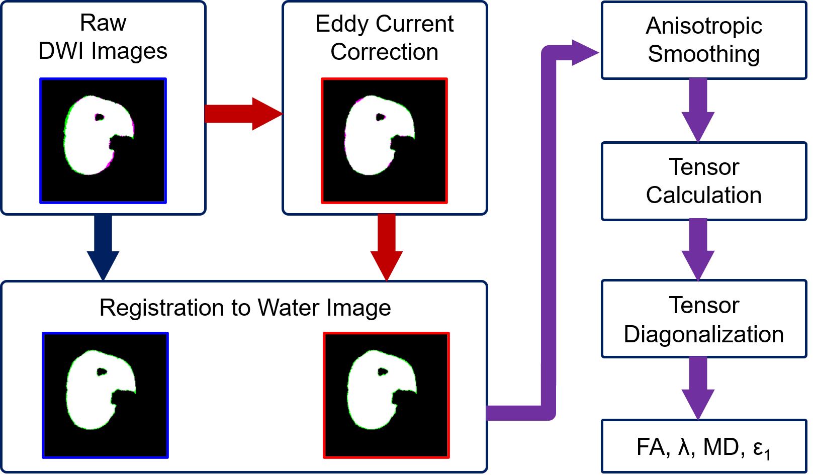
Figure 1. General workflow of data analysis. Mask difference between
diffusion-weighted images and water images are shown. Eddy current improves the
morphological match between diffusion-weighted and water images.
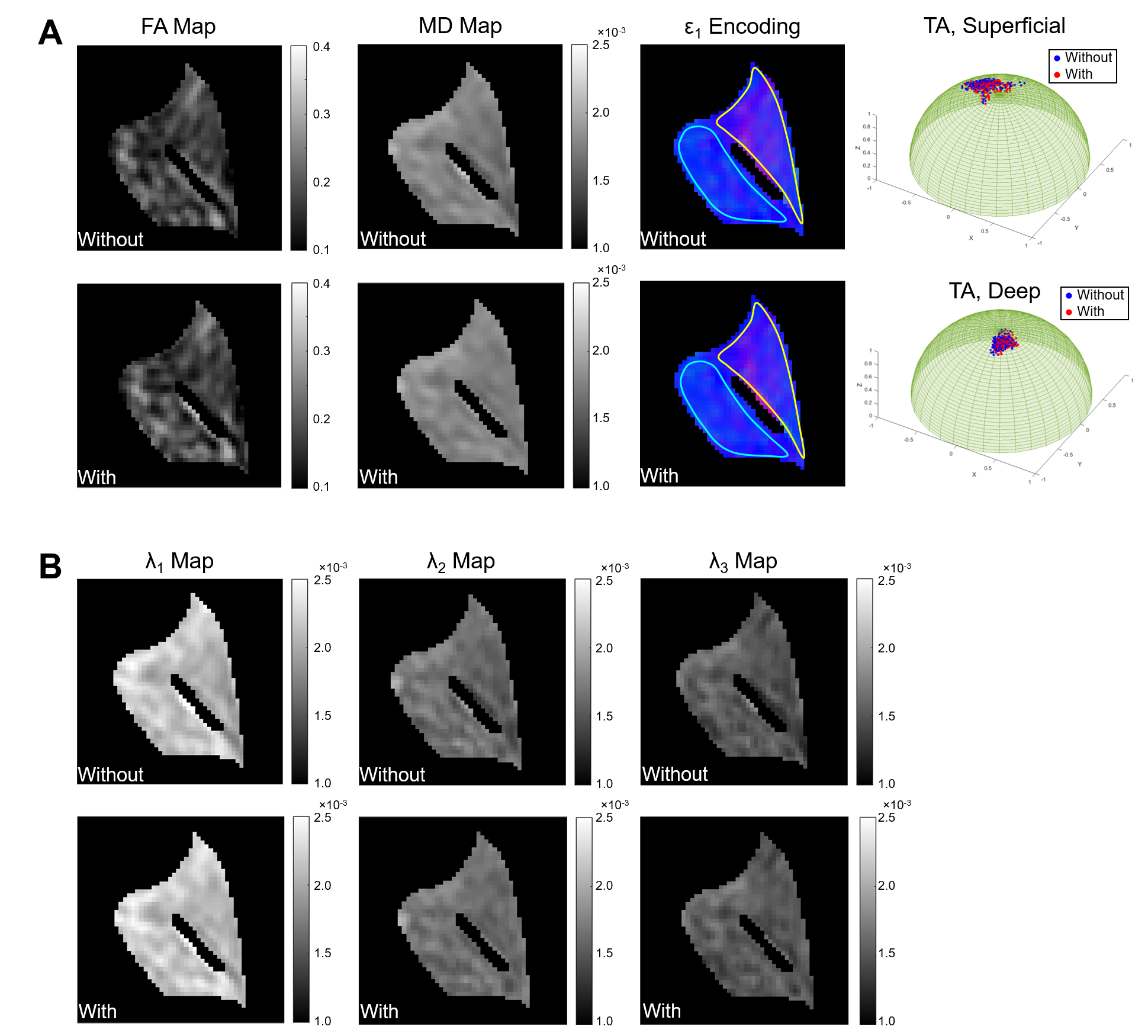
Figure 2. Example of comparison of derived indices from muscle diffusion tensor
with and without eddy current correction in tibialis anterior (TA) muscle. (A)
Comparison of FA map, MD map, color-encoded map of the first eigenvector (ε1), and projection of ε1
on unit hemisphere within the superficial and deep compartments of TA. Color
scheme of direction encoding: red: x-direction, green: y-direction,
blue: z-direction; superficial and deep compartments of the TA muscle
are delineated by yellow and aqua boundaries in ε1 maps,
respectively. (B) Comparison of map of eigenvalues.