Kihwan Kim1, Yuning Gu1, Yuran Zhu1, Yudu Li2,3, Sherry Huang1, Zhi-Pei Liang 2,3, and Xin Yu1,4
1Biomedical Engineering, Case Western Reserve University, cleveland, OH, United States, 2Electrical and Computer Engineering, University of Illinois at Urbana-Champaign, Urbana, IL, United States, 3Beckman Institute for advanced Science and Technology, University of Illinois at Urbana-Champaign, Urbana, IL, United States, 4Case Center for Imaging Research, Case Western Reserve University, cleveland, OH, United States
1Biomedical Engineering, Case Western Reserve University, cleveland, OH, United States, 2Electrical and Computer Engineering, University of Illinois at Urbana-Champaign, Urbana, IL, United States, 3Beckman Institute for advanced Science and Technology, University of Illinois at Urbana-Champaign, Urbana, IL, United States, 4Case Center for Imaging Research, Case Western Reserve University, cleveland, OH, United States
31P-MRS was used to
evaluate changes in muscle metabolism in a mouse model of Duchenne muscular
dystrophy, the mdx mouse. We observed altered
creatine kinase activity and phosphocreatine recovery rate, suggesting abnormal mitochondrial function in the skeletal muscle of mdx mice.
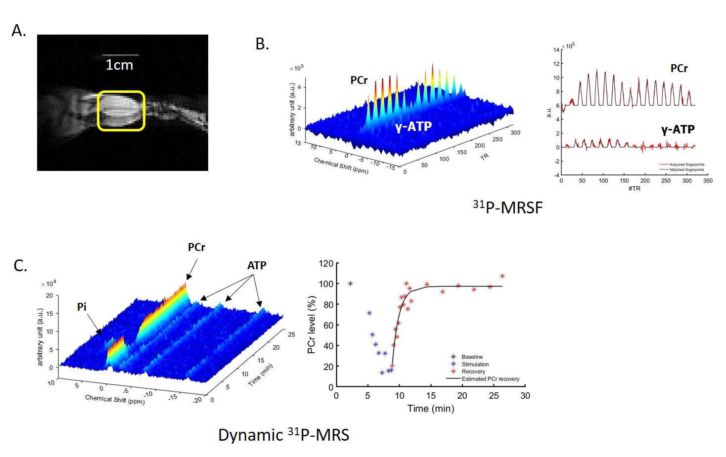
Figure 1. (A) Coronal image of mouse
hindlimb with a 1cm long landmark to display the length and location of 31P-
saddle coil; (B) 3D 31P-MR
fingerprints and 2D 31P-MR fingerprints matched to a dictionary
entry (C) 3D and 2D dynamic 31P-MR
spectra
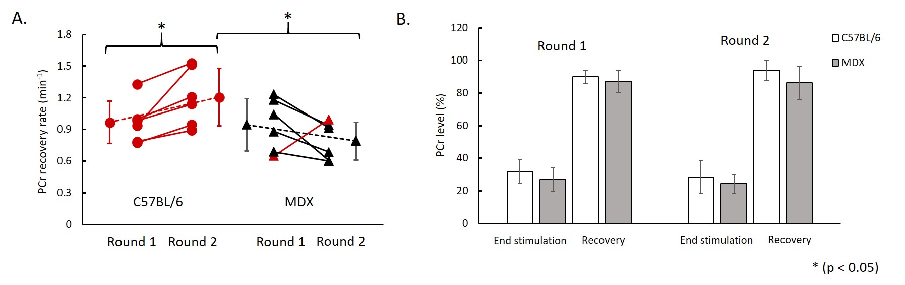
Figure 3. (A) PCr
recovery rate between round 1 and round 2 stimulations (red color indicates increase in PCr recovery
rate and black color indicates decrease in PCr recovery rate); (B) PCr level at
the end of stimulation-induced muscle contraction and at after 16 min recovery.