Natenael B Semmineh1, Sudarshan Ragunathan2, Laura C Bell2, and C Chad Quarles 2
1Imaging Research, Barrow Neurological Institute rrow Neurological Institute, Phoenix, AZ, United States, 2Imaging Research, Barrow Neurological Institute, Phoenix, AZ, United States
1Imaging Research, Barrow Neurological Institute rrow Neurological Institute, Phoenix, AZ, United States, 2Imaging Research, Barrow Neurological Institute, Phoenix, AZ, United States
Tissue transverse relaxivity at tracer equilibrium (TRATE) can longitudinally differentiate myofiber atrophy in an ALS model.
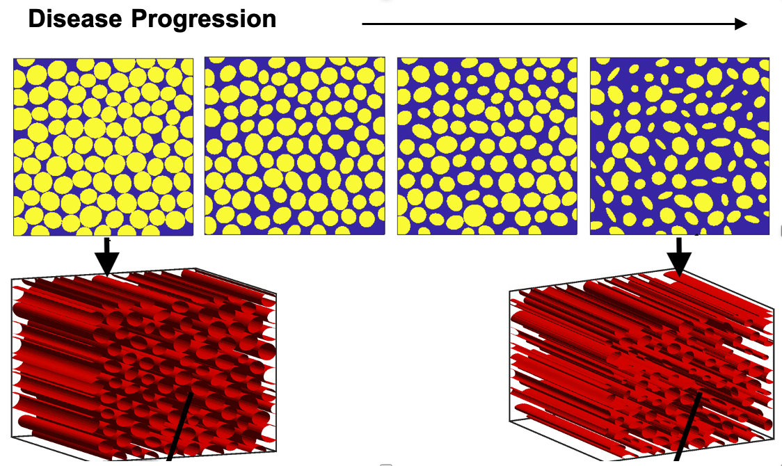
Figure 1: Representative tissue structure that will be used to characterize RCI in silico, as a function of ALS progression. Top row shows a 2D section of the 3D structure below.
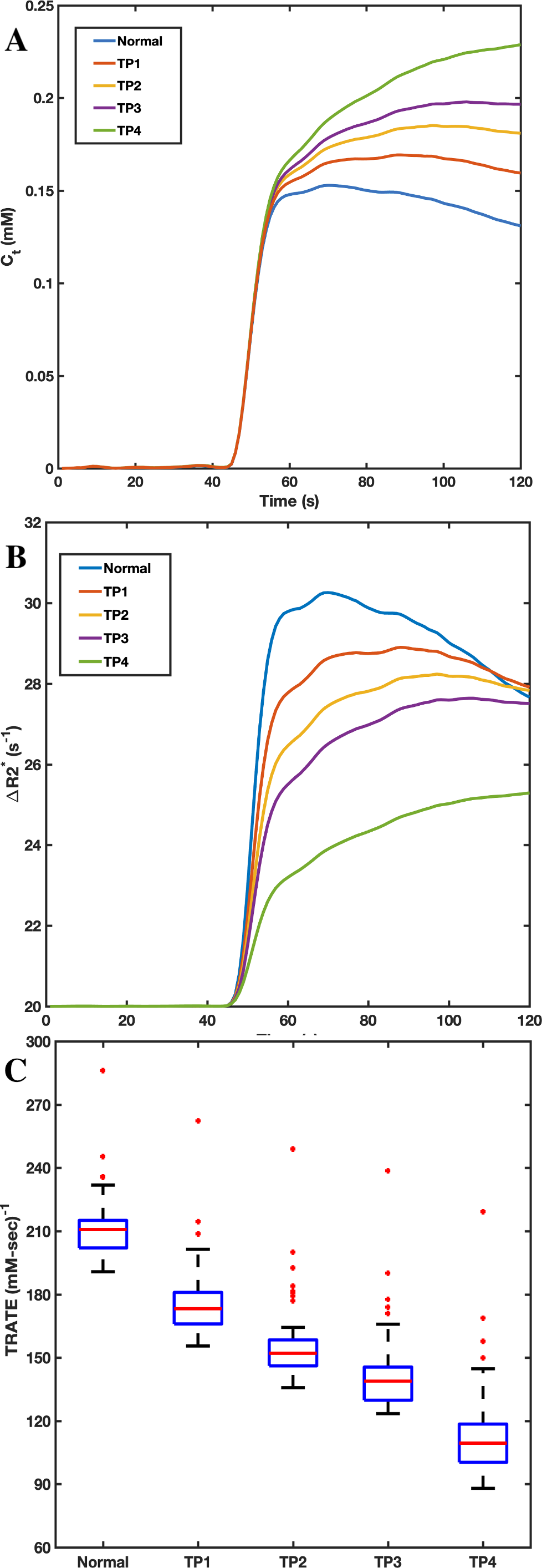
Figure 2: Example concentration time curves (A), ΔR2* time curves (B) and the corresponding TRATE values (C) over four diseases progression time points (TP) along with a normal myofiber case. Note each profile in ALS are dissimilar in shape and magnitude both for ΔR2* and concentration.