Matthew Birkbeck1,2,3, Linda Heskamp1, Ian Schofield1, Roger Whittaker1, and Andrew Blamire1
1Translational and Clinical Research Unit, Newcastle University, Newcastle upon Tyne, United Kingdom, 2Newcastle Biomedical Research Centre, Newcastle Biomedical Research Centre, Newcastle upon Tyne, United Kingdom, 3Northern Medical Physics and Clinical Engineering, Newcastle upon Tyne NHS Foundation Trust, Newcastle upon Tyne, United Kingdom
1Translational and Clinical Research Unit, Newcastle University, Newcastle upon Tyne, United Kingdom, 2Newcastle Biomedical Research Centre, Newcastle Biomedical Research Centre, Newcastle upon Tyne, United Kingdom, 3Northern Medical Physics and Clinical Engineering, Newcastle upon Tyne NHS Foundation Trust, Newcastle upon Tyne, United Kingdom
MUMRI has been applied for the
first time to study in-vivo single human MUs in the forearm and hand muscles
and to determine the size and shapes of these MUs. This increases the clinical
translatability of the MUMRI technique.
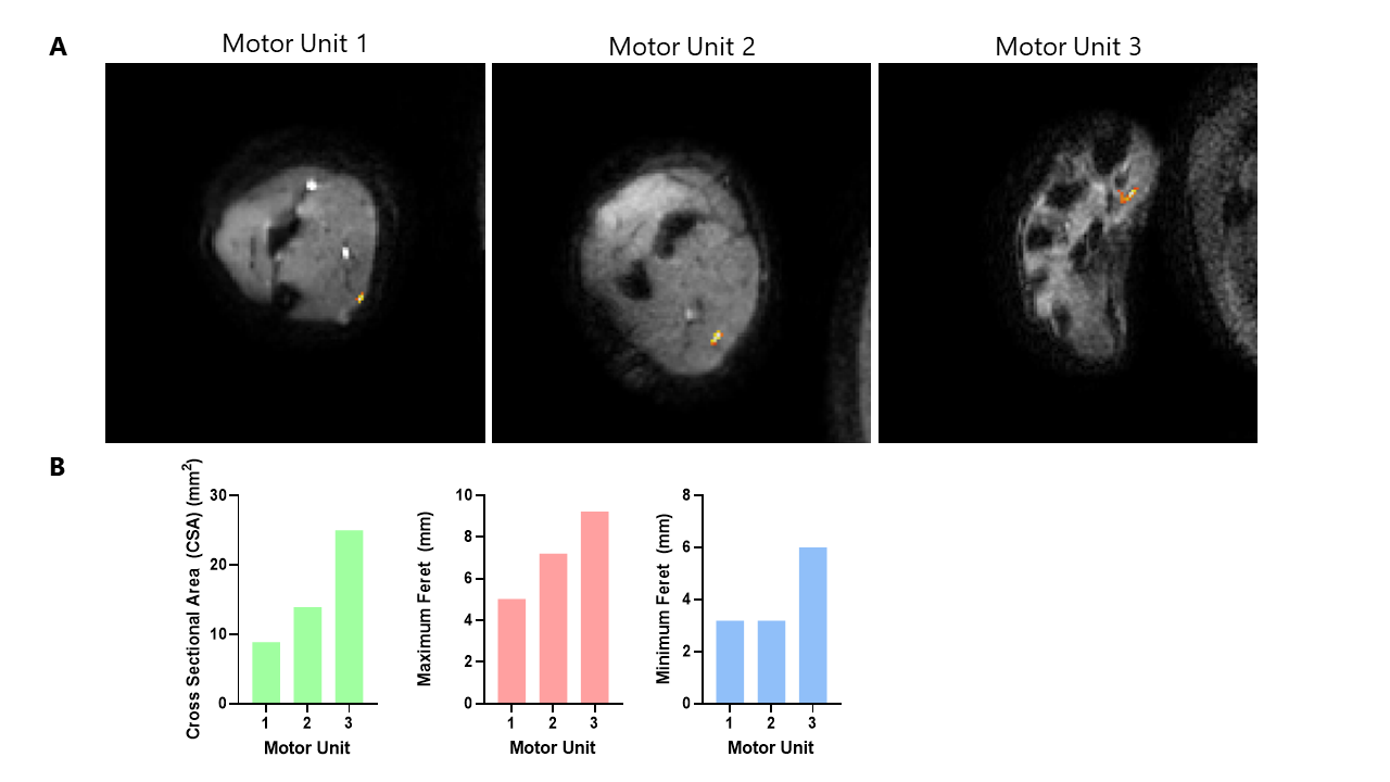
Figure
4: A) MU difference maps overlaid onto the MUMRI
images for each of the extracted MUs. Two from the forearm in the flexor carpi
ulnaris muscle and one from the hand in the abductor pollicis muscle. B) Bar
charts showing MU metrics: cross sectional area (CSA) shown in green, maximum
Feret diameter shown in red and minimum Feret diameter shown in blue.
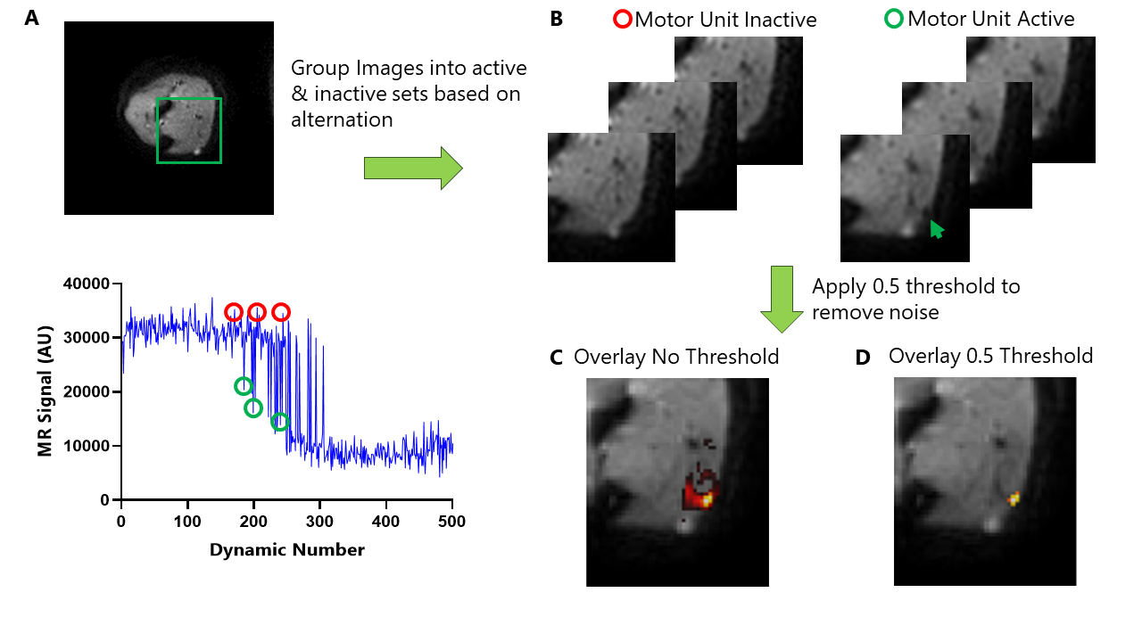
Figure
2: Data processing
steps to extract the single MUs. A) MUMRI image B) Images grouped into two data
sets: where motor unit is inactive and active (indicated by red arrow). Graph
shows single voxel time-series from the MU territory, example alternations
where MU inactive (red circle), MU active (green circles). C) MU difference map
with no threshold applied. D) MU difference map with 0.5 threshold, showing
good agreement with initial size of MU territory in (B).