Hon J Yu1, Saya Horiuchi1,2, Toshimi Tando1, Vincent J Caiozzo3, Virginia E Kimonis4, and Hiroshi Yoshioka1
1Radiological Sciences, University of California, Irvine, Orange, CA, United States, 2Radiology, St. Luke's International Hospital, Tokyo, Japan, 3Department of Orthopaedics, Physiology & Biophysics, University of California, Irvine, Irvine, CA, United States, 4Division of Genetic and Genomic Medicine, Department of Pediatrics, University of California, Irvine, Irvine, CA, United States
1Radiological Sciences, University of California, Irvine, Orange, CA, United States, 2Radiology, St. Luke's International Hospital, Tokyo, Japan, 3Department of Orthopaedics, Physiology & Biophysics, University of California, Irvine, Irvine, CA, United States, 4Division of Genetic and Genomic Medicine, Department of Pediatrics, University of California, Irvine, Irvine, CA, United States
Texture features can
classify varying degrees of muscle myopathy when properly trained in supervised
machine-learning framework.
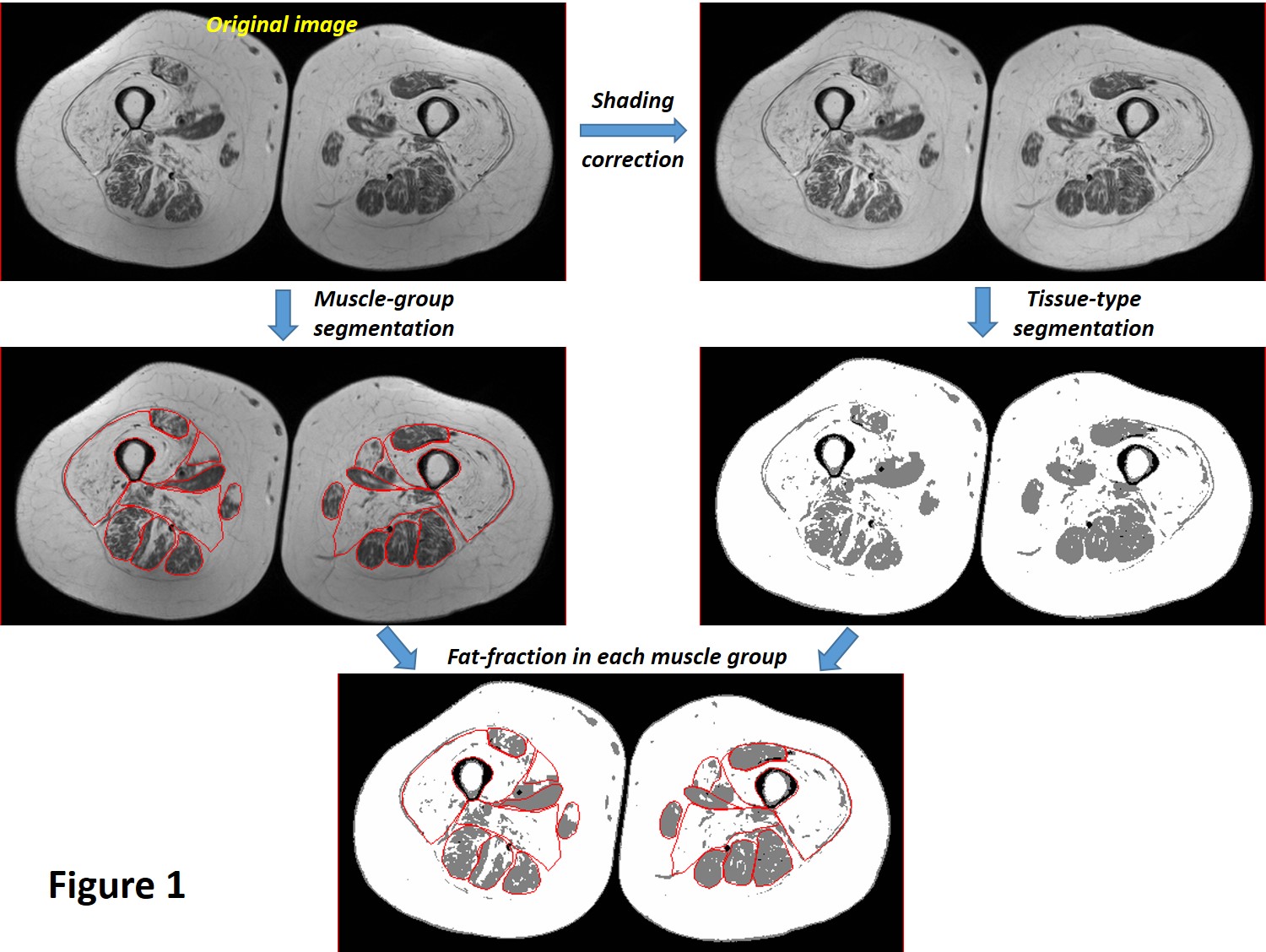
The
overall workflow of fat-fraction calculation that utilizes manual muscle
segmentation, shading/intensity-correction across FOV, and classification of
different tissue types based on a 3-class fuzzy c-means (FCM) algorithm.
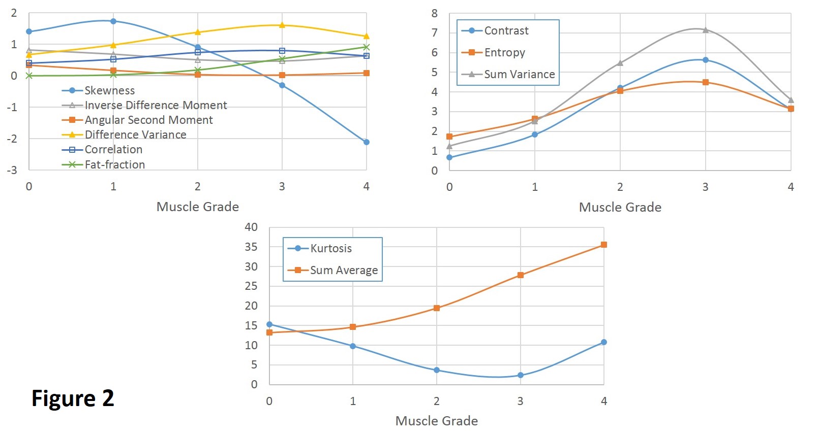
Plots
of group-averaged fat-fraction and texture parameter values with similar
vertical scales in order to better visualize their trends as a function of
muscle grades.