Arthur Jourdan1, Arnaud Le Troter2, Pierre Daude2, Stanislas Rapacchi2, Catherine Masson1, Thierry Bege1,3, and David Bendahan2
1Aix-Marseille Univ, Univ Gustave Eiffel, IFSTTAR, LBA, Marseille, France, 2Aix Marseille Univ, CNRS, CRMBM, Marseille, France, 3Department of General Surgery, Aix Marseille Univ, North Hospital, APHM, Marseille, France
1Aix-Marseille Univ, Univ Gustave Eiffel, IFSTTAR, LBA, Marseille, France, 2Aix Marseille Univ, CNRS, CRMBM, Marseille, France, 3Department of General Surgery, Aix Marseille Univ, North Hospital, APHM, Marseille, France
A novel semi-automatic post-processing method dedicated to real-time dynamic MRI aiming at a fast and reliable quantification of abdominal wall muscles deformations based on a supervised 2D+t segmentation (mean Dice similarity coefficient of 0.95), masks registration and parcellation.
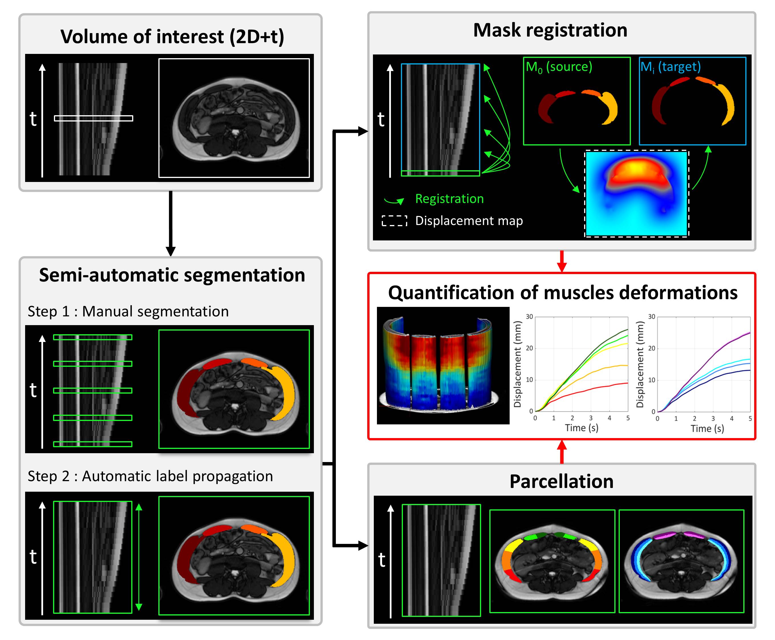
Figure 2 : Quantification
of muscles deformations pipeline; A : 2D+t volume of interest in sagittal view
with an arbitrary selected axial slice.
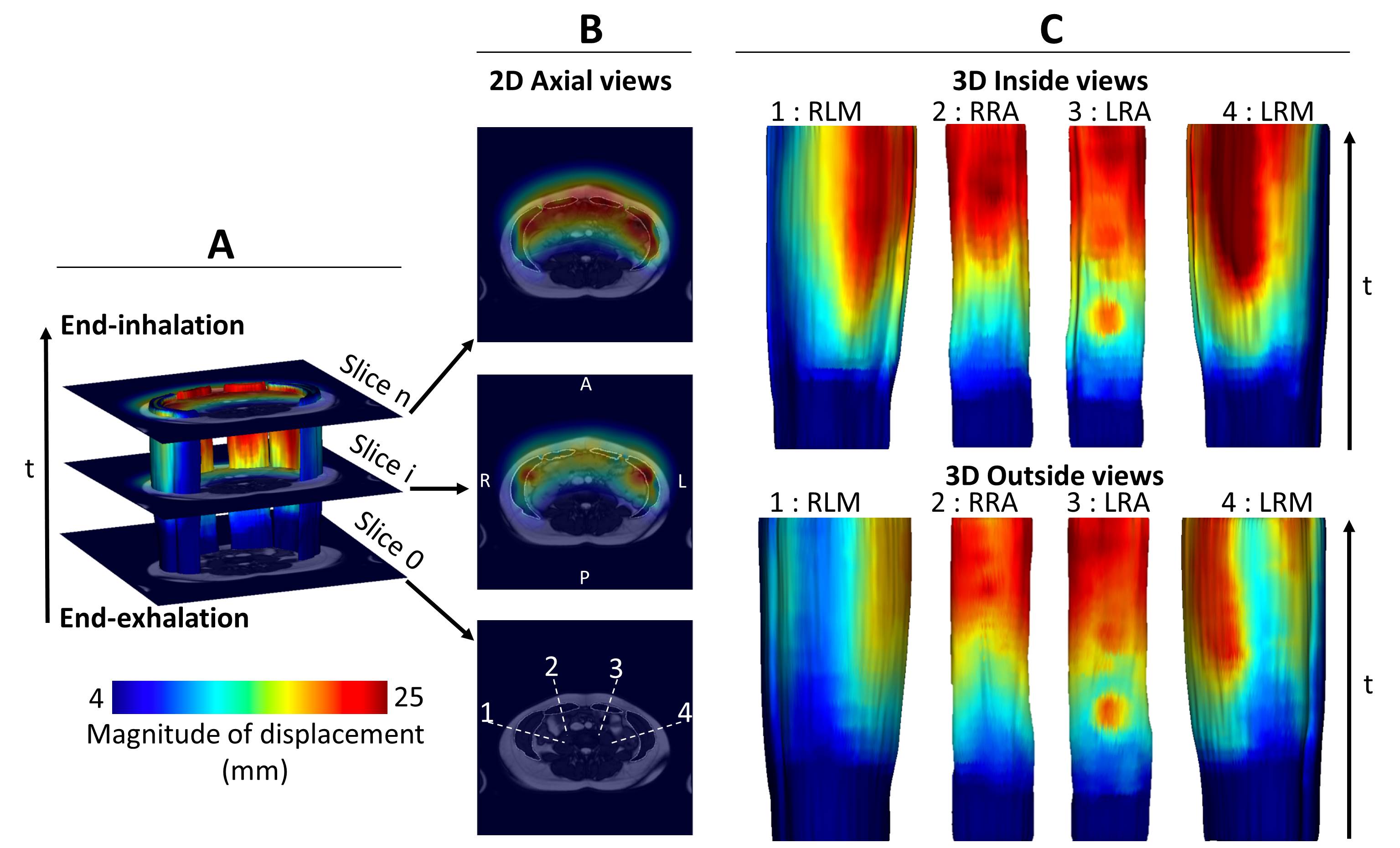
Figure 3 : Typical
magnitude of displacement maps. These maps were obtained for the subject
closest to the mean values of the cohort; A : 3d general view of displacement
magnitude within the abdominal wall muscles from end-exhalation to
end-inhalation with three anatomical MRI slices; B : 2D axial views of the
magnitude of displacement computed on the three previous anatomical MRI slices;
C : 3D visualization of the magnitudes within each individual muscle group.