Cyril Tous, PhD1, Alexandre Jodoin, MD2, Detlev Grabs, MD, PhD3, Elijah Van Houten, PhD4, and Nathalie J Bureau, MD MSc FRCP(C)1,2
1Radiology, Centre de recherche du Centre hospitalier de l’Université de Montréal, Montreal, QC, Canada, 2Radiology, Centre hospitalier de l’Université de Montréal, Montréal, QC, Canada, 3Anatomy, Université du Québec à Trois-Rivières, Trois-Rivières, QC, Canada, 4Mechanical Engineering, Université de Sherbrooke, Sherbrooke, QC, Canada
1Radiology, Centre de recherche du Centre hospitalier de l’Université de Montréal, Montreal, QC, Canada, 2Radiology, Centre hospitalier de l’Université de Montréal, Montréal, QC, Canada, 3Anatomy, Université du Québec à Trois-Rivières, Trois-Rivières, QC, Canada, 4Mechanical Engineering, Université de Sherbrooke, Sherbrooke, QC, Canada
Rotator cuff tears lead to fatty infiltration and fibrosis,
causing stiffness and decreased elasticity. Diffusion Tensor Imaging
stabilizes regularization in elastography. Good DTI repeatability is achieved in
six asymptomatic volunteers and myocytes’ tracks are retrieved.
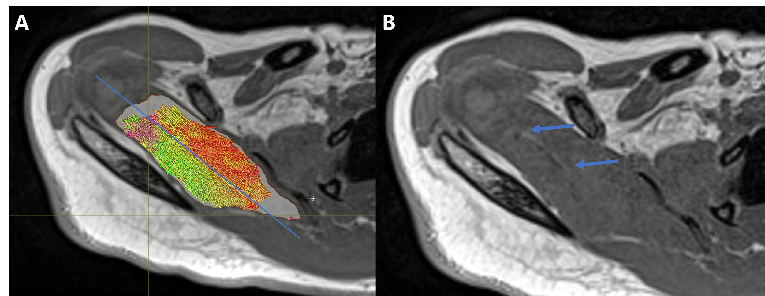
Figure 4) The
mask delineates the supraspinatus (A-white background on muscle) where
tractography is performed (A-colors) overlaid by a T1 VIBE image. The line separation (A-blue line) corresponds
to the separation between the posterior and anterior bundle (B-arrows) where myocytes insert posteriorly (red
tracks) and anteriorly (green track).

Figure 2) Bland-Altman
plots of FA across three scans (pairwise comparison of scans 1-2, 1-3, 2-3) for
each volunteer (1 to 6) in the supraspinatus (sup) or infraspinatus (inf).
Limit of agreement (loa) is 0.071 with a bias at 0.0.