Shuai Zhang1, Xiangtao Lin2, Ximing Wang2, Xiang Feng3, and Rui Diao2
1School of Medicine, Shandong First Medical University, School of Medicine, Shandong First Medical University, Jinan, China, 2Department of Radiology, Shandong Provincial Hospital affiliated to Shandong First Medical University, Jinan, China, 3MR Scientific Marketing, Diagnosis Imaging, Siemens Healthcare Ltd, Beijing, China
1School of Medicine, Shandong First Medical University, School of Medicine, Shandong First Medical University, Jinan, China, 2Department of Radiology, Shandong Provincial Hospital affiliated to Shandong First Medical University, Jinan, China, 3MR Scientific Marketing, Diagnosis Imaging, Siemens Healthcare Ltd, Beijing, China
The cervical, thoracic, lumbar, and L1–L5 centrum ossification centers show good correlation with gestational age in the second and third trimesters. The L1 centrum ossification center is best suited as a marker for fetal cervical, thoracic, and lumbar development.
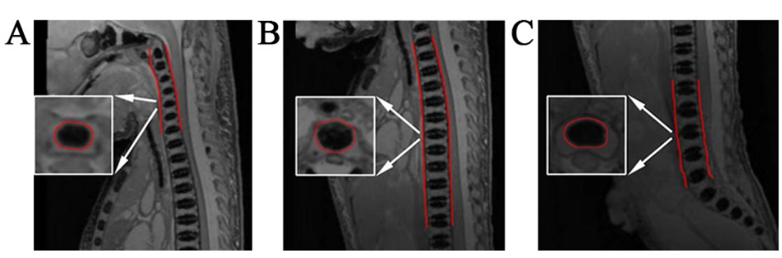
Fig 1. Sample segmentations of cervical (A), thoracic (B), and lumbar (C) centrum ossification center volumes.
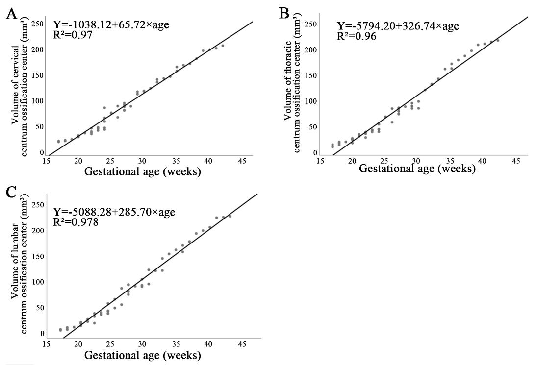
Fig 2. Regression lines for the volumes of the cervical (A), thoracic (B), and lumbar (C) centrum ossification centers.