1Radiology, Peking University Third Hospital, Beijing, China, 2Center for Functional Onco-Imaging, Irvine, CA, United States, 3Radiology, Peking University International Hospital, Beijing, China
For patients with spinal GCTB, preoperative MRI observed multiple cystic changes and vertebral compression≥ 50%, suggesting a poor prognosis. The expression of vascular endothelial growth factor and p53 tumor suppressor gene may not be associated with postoperative recurrence.
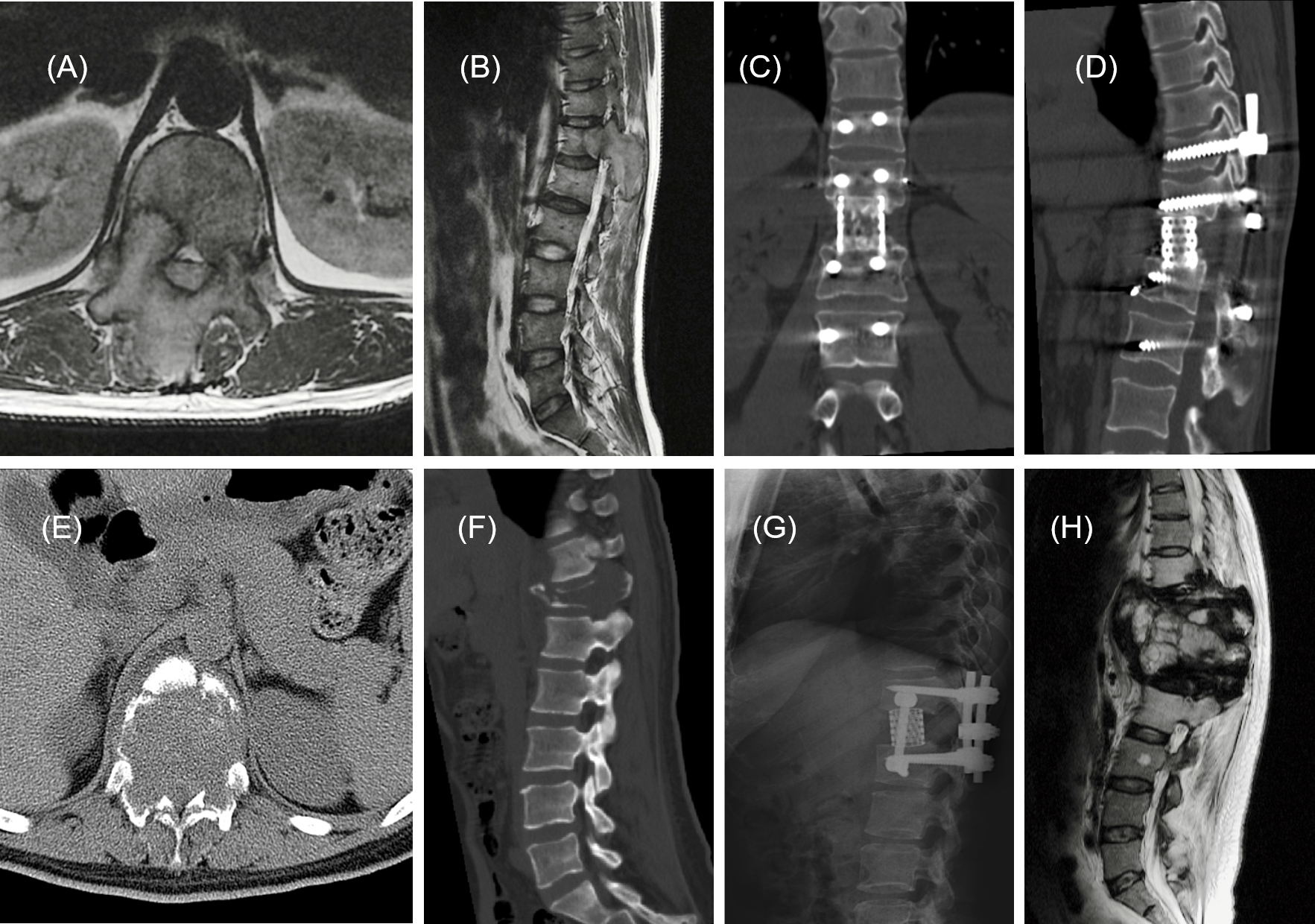
Fig.2. Top panel: A 35-year-old man,(A) and (B): MR images showed a mass on the T12 vertebra, bilateral pedicle and lamina with extension into the spinal canal, managed with en bloc resection (C), at a 36-month follow-up review, there was no evidence of recurrence (D), and now the patient is still on visit.
Bottom panel: A 35-year-old woman, maximum diameter of lesion is 55mm (E),with pathologic fracture of the T12 vertebra (F) , managed with en bloc resection (G). The sagittal T2-WI MR image at 12-month follow-up, recurrence was detected (H), and confirmed by pathology with puncture.