Amir Masoud Afsahi1, Zhao Wei1, Michael Carl2, Saeed Jerban1, Hyungseok Jang1, Nicole Le1, Jiang Du1, Eric Y. Chang1,3, and Ya-Jun Ma1
1Department of Radiology, University of California, San Diego, San Diego, CA, United States, 2GE HealthCare, San Diego, CA, United States, 3Radiology Service, Veterans Affairs, San Diego Healthcare System, San Diego, CA, United States
1Department of Radiology, University of California, San Diego, San Diego, CA, United States, 2GE HealthCare, San Diego, CA, United States, 3Radiology Service, Veterans Affairs, San Diego Healthcare System, San Diego, CA, United States
By 3D slab-selective UTE MRI, we are able to have a high resolution in lumbar spinal bone comparable to CT as gold standard. This is specially important when we need images in patients more sensitive to X-Ray exposure like children, pregnant women or anybody who needs periodic imaging.
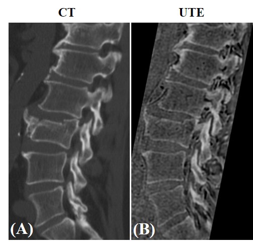
Figure 4. CT (A) and
UTE (B) lumbar spinal bone images from 72-year-old
male patient with spinal fractures. Bone fractures can be seen clearly in both
CT and UTE images.
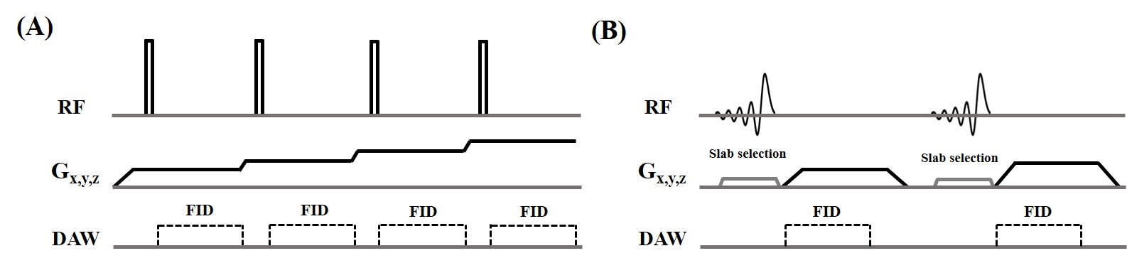
Figure 1. 3D ZTE (A) and UTE (B) sequences. The ZTE sequence utilizes a non-selective rectangular RF pulse with short duration for excitation, followed by 3D center-out radial sampling. To minimize echo time, readout gradients are turned on prior to signal excitation so that the gradient encoding can begin simultaneously with signal excitation. The 3D UTE sequence enables slab selection by using a soft-half pulse for excitation together with a slice-selective gradient. After excitation, the spatial encoding gradient is turned on and simultaneous data acquisition begins.