Kaiwen Yang1, Baofa Luo1, Yilong Huang1, Lisha Nie2, and Bo He1
1Medical imaging department, The First Affiliated Hospital of Kunming Medical University, Kunming, China, 2GE Healthcare,MR Research China, Beijing, China
1Medical imaging department, The First Affiliated Hospital of Kunming Medical University, Kunming, China, 2GE Healthcare,MR Research China, Beijing, China
Based on functional MRI, this study
explored the influence mechanism of rehabilitation exercise on rat paraspinal
muscles and intervertebral discs. It is found that rehabilitation exercise can
reduce TNF-α content and improve paravertebral muscular atrophy.
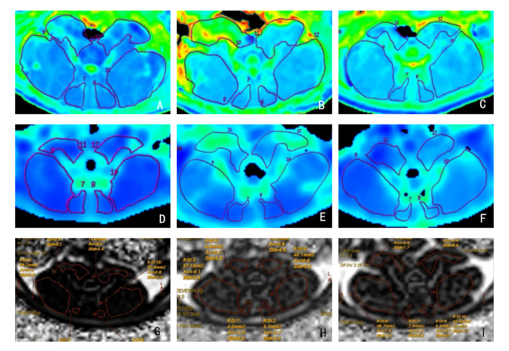
Figure.3 Color-coded T2-calculated, R2* and fat fraction maps. In T2-calculated maps, blue represents areas of short T2 values. T2 maps of normal (A), DLBP(B) and swimming group (C). In R2* maps, blue represents areas of lower R2* value. R2* maps of normal (D), DLBP group(E) and swimming group (F). In fat fraction maps, white represents areas of higher fat fraction. Fat fraction maps of normal (G), DLBP(H) and swimming group(I).
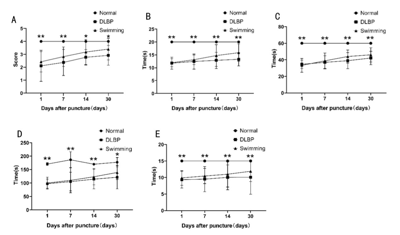
Figure.1 Behavioral test results of three groups within one month after operation. (A)Gait test; (B) hot-plate test; (C)acetone test; (D)tail suspension test; (E)grip strength test. Data are reported as mean ± standard deviation of mean; *p<0.05, **P<0.01(Kruskal-Wallis test).