Yongye Chen1, Enlong Zhang 2, Qizheng Wang 1, Huishu Yuan1, Huishu Yuan1, Hongqing Zhuang1, and Ning Lang1
1Peking University Third Hospital, Beijing, China, 2Peking University International Hospital, Beijing, China
1Peking University Third Hospital, Beijing, China, 2Peking University International Hospital, Beijing, China
DCE-MRI can
be used to evaluate local tumor response. In our study, Ktrans, Kep
and Ve were found to be of great value in evaluating the efficacy of
CyberKnife radiosurgery in spinal metastases. ΔKtrans had the highest
diagnostic efficiency, with an AUC of 0.821.
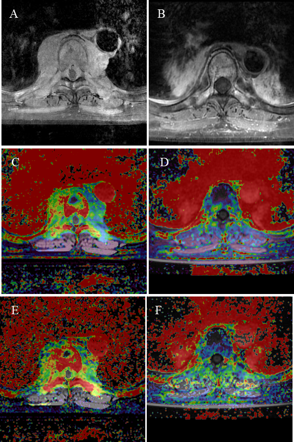
One case in the non-PD group. (A): Axial
T1WI-enhanced MRI showed a huge soft tissue mass around the vertebra before
treatment; (B): Three months after treatment, axial T1WI enhanced MRI showed
the lesion soft tissue mass was significantly reduced; (C): Pretreatment Ktrans
was 0.342 min-1; (D): Post-treatment Ktrans was 0.121 min-1,
decreased by 64.6%; (E): Pretreatment Kep was 2.445 min-1;
(F): Post-treatment Kep was 0.959 min-1, decreased by 60.8%.

One case in PD group. (A): Enhanced T1WI scan
axial view showed a soft tissue mass invading the spinal canal on the left side
of the vertebra, and the spinal cord was compressed; (B): Axial T1WI enhanced
image showed that the soft tissue mass of the lesion was slightly larger than
before, and the low signal cystic area is seen behind the spinal canal; (C): Pretreatment
Ktrans was 0.503 min-1; (D): Post-treatment Ktrans
was 0.750 min-1, increased by 49.1%; (E): Pretreatment Kep was
2.487 min-1; (F): Post-treatment Kep was 4.187 min-1,
increased by 68.4%.