Zhilin Ji1, Weiqiang Dou2, Yuefen Zou1, Yin Shi1, Yu Zheng1, and Hongyuan Ding1
1The First Affiliated Hospital of Nanjing Medical University, Nanjing, China, 2GE Healthcare, MR Research China, Beijing, P.R. China, Beijing, China
1The First Affiliated Hospital of Nanjing Medical University, Nanjing, China, 2GE Healthcare, MR Research China, Beijing, P.R. China, Beijing, China
we aimed to investigate UTE's feasibility for assessing the CEP damage and evaluate the relationship between the CEPs’ grading and intervertebral disc degeneration. Strong correlation was observed between CEP and intervertebral disc degeneration. So, UTE can assist to assess the CEP damage.
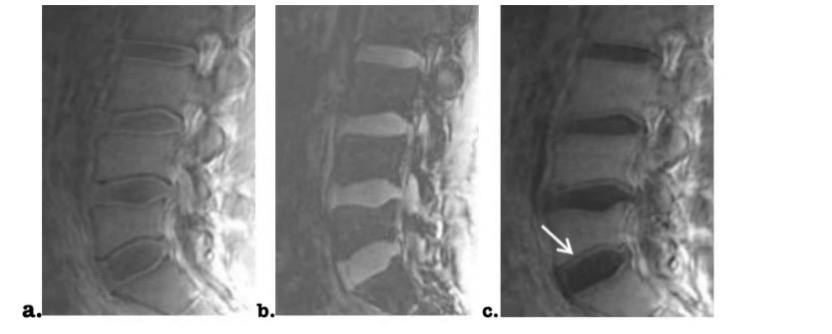
Fig2. Subtracting a 3D ultrashort Echo Time (UTE) image acquired at short TE of 0.03 ms (a) by the one acquired at long TE of 6 ms (b), a resultant subtracted 3D UTE image (c) of a lumbar spine with relatively normal cartilage endplate (CEP) is presented at sagittal view. Well structural CEP (denoted by white arrow) is hyperintense as well as adjacent hypointense vertebral endplate. The effect of nucleus pulposus, annulus fibrosus was removed after image subtraction.
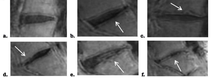
Fig3. On the subtracted 3D UTE images between short and long TEs, cartilage endplates (CEPs) were classified into six grades. Representative CEP images at each grade are shown (a: type I, b: type II, c: type III, d: type IV, e: type V, f: type VI ).