Xing Yin1, Xin Zhao1, Liying Zhang1, Qingna Xing1, Rui Yuan2, and Zhijun Niu3
1Radiology, the Third Affiliated Hospital of Zhengzhou University, Zhengzhou, China, 2Ultrasound, the Third Affiliated Hospital of Zhengzhou University, Zhengzhou, China, 3Obstetrics and Gynecology, the Third Affiliated Hospital of Zhengzhou University, Zhengzhou, China
1Radiology, the Third Affiliated Hospital of Zhengzhou University, Zhengzhou, China, 2Ultrasound, the Third Affiliated Hospital of Zhengzhou University, Zhengzhou, China, 3Obstetrics and Gynecology, the Third Affiliated Hospital of Zhengzhou University, Zhengzhou, China
There are good linear correlations between the
heights of LVBOC and GA,
lengths of LVBOC and GA,
heights of intervertebral gap and GA.The LVBOCs may show linear T2 hyperintensity in
the mid-sagittal plane (14/465 vertebra). The positions of conus
level lies from L1 to L3.

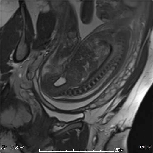
Figure
1 24 gestational weeks. Figure 1A, measuring of height
and length of L5 vertebral ossification body center, and height of L3-4 IVP.
Figure 1B, the location of conus medullaris.
Figure
2 24 gestational weeks, the mid-sagittal plane of lumbar
spine showed linear T2 hyperintensity in L2 and L4 vertebral ossification body
center.