Fangfang Fu1, Nan Meng2, Zhun Huang3, Yaping Wu1, Pengyang Feng3, Xiaochen Li1, Yan Bai1, Wei Wei1, Jianmin Yuan4, Tianyi Xu4, and Meiyun Wang1
1Department of Medical Imaging, Henan Provincial People’s Hospital, People’s Hospital of Zhengzhou University, Zhengzhou, China, 2Department of Radiology, Zhengzhou University People’s Hospital & Henan Provincial People’s Hospital, Academy of Medical Sciences, Zhengzhou, China, 3Department of Radiology, Henan University People’s Hospital & Henan Provincial People’s Hospital, School of Basic Medicine. Department of Radiology, Zhengzhou University People’s Hospital & Henan Provincial People’s Hospital, Academy of Medical Sciences., Zhengzhou, China, 42258 Chengbei Road, Jiading District, Shanghai, China 201907, Shanghai, China
1Department of Medical Imaging, Henan Provincial People’s Hospital, People’s Hospital of Zhengzhou University, Zhengzhou, China, 2Department of Radiology, Zhengzhou University People’s Hospital & Henan Provincial People’s Hospital, Academy of Medical Sciences, Zhengzhou, China, 3Department of Radiology, Henan University People’s Hospital & Henan Provincial People’s Hospital, School of Basic Medicine. Department of Radiology, Zhengzhou University People’s Hospital & Henan Provincial People’s Hospital, Academy of Medical Sciences., Zhengzhou, China, 42258 Chengbei Road, Jiading District, Shanghai, China 201907, Shanghai, China
Multiparametric PET/MRI is potentially useful
in distinguishing malignant and benign pulmonary lesions. The
combination of several IVIM parameters and PET parameters could improve the differentiation of malignant and benign pulmonary lesions.
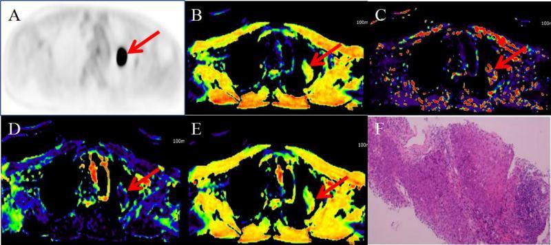
Figure 1.A 61-year-old man with left lung squamous cell
carcinoma. The red arrows point to
the lesion. A. PET image. B. Dt
pseudo colored map. C. Dp pseudo colored map.
D. F pseudo colored maps. E. sADC pseudo colored map. F. Hematoxylin and
eosin(HE)x 100.
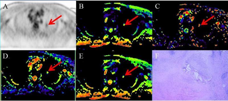
Figure 2.A 56-year-old man with
left lung tuberculous granuloma. The red arrows point to the lesion.A. PET
image. B. Dt pseudo colored map. C. Dp pseudo colored
map. D. F pseudo colored maps. E. sADC pseudo colored map. F. Hematoxylin and
eosin(HE)x 100.