Gaofeng Shi1, Liyun Zheng2, Yongming Dai2, Hui Liu1, Hui Feng1, and Hongshan Zhu1
1Department of Radiology, Fourth Hospital of Hebei Medical University, Shijiazhuang, China, 2United Imaging Healthcare, Shanghai, China
1Department of Radiology, Fourth Hospital of Hebei Medical University, Shijiazhuang, China, 2United Imaging Healthcare, Shanghai, China
This
study suggested that the combined measurement of OE-UTE-MRI and IVIM-DWI may
serve as a promising method for the noninvasive assessment of lung function and
classification of LUAD subtype.
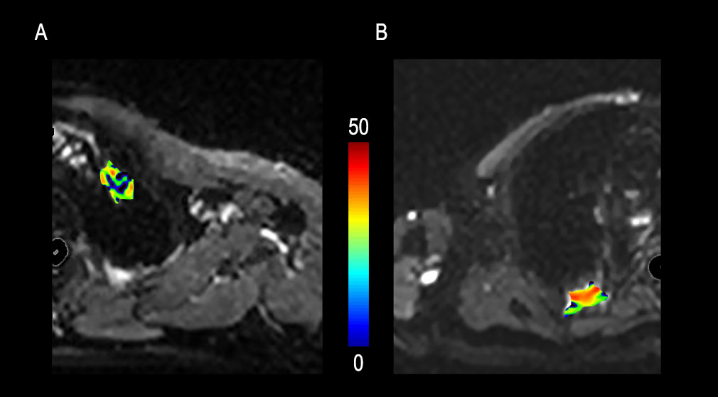
Figure 3. Representative IVIM-DWI analysis for lepidic predominant adenocarcinoma and micropapillary predominant adenocarcinoma. (A) f map from a lepidic predominant adenocarcinoma patient; (B) f map from a micropapillary predominant adenocarcinoma patient.

Figure 4. Representative OE-UTE-MRI analysis for lepidic predominant adenocarcinoma and micropapillary predominant adenocarcinoma. (A) lesion PSE map from a lepidic predominant adenocarcinoma patient; (B) lesion PSE map from a micropapillary predominant adenocarcinoma patient.