Dandan Peng1, Cong Xia1, Yuancheng Wang1, Zhongshuai Zhang2, and Shenghong Ju1
1Zhongda Hospital, Medical School of Southeast University, Nanjing, China, 2SIEMENS Healcare, Shanghai, China
1Zhongda Hospital, Medical School of Southeast University, Nanjing, China, 2SIEMENS Healcare, Shanghai, China
This study is expected to provide
information about differentiating of benign from malignant pulmonary lesions
with GRASP-DCE MRI among
33 lung lesions. The results initially indicated that the quantitative DCE
parameters could be a useful technique
for lung lesions detection.
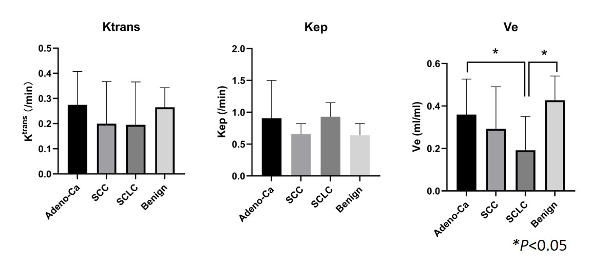
Figure1.The GRASP DCE-derived quantitative
values distribution of malignancy and benignity in lung lesions. The mean
values of Ve in Adeno-Ca and benign groups are 0.360±0.167 and 0.428±0.114. (ANOVA,
p<0.05)
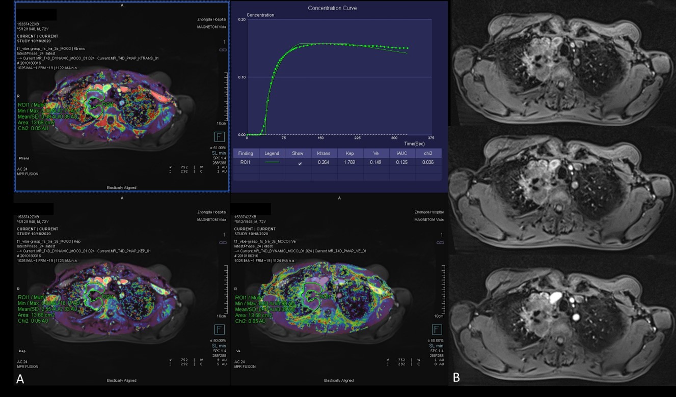
Figure 3. A
72-year-old male with a 6cm mass in the right upper lobe. This mass was
diagnosed as adenocarcinoma IIIb (cT4N2M0). (A): Free-breathing
Golden-angle RAdial Sparse Parallel (GRASP)-DCE MRI post-processed image
showing mean Ktrans of 0.264 min-1, Kep of 1.769 min-1 and Ve of 0.149. The iAUC
value of Time-intensity curve for this mass was 0.125. (B):There
were 3 early phases images from top to the bottom after contrast injection
showing the mass continuous heterogeneous enhancement.