Fu Yicheng1, Zhang Zhongshuai2, Yu Ye1, and Wu Huawei1
1Radiology, Renji Hospital, School of Medicine, Shanghai Jiaotong University, Shanghai, China, 2SIEMENS Healthcare, China, Shanghai, China
1Radiology, Renji Hospital, School of Medicine, Shanghai Jiaotong University, Shanghai, China, 2SIEMENS Healthcare, China, Shanghai, China
Quantitative T2 mapping and DW-IVIM parameters could
provide valuable information and serve as a supplementary imaging marker for
differentiating malignant from benign SPNs.
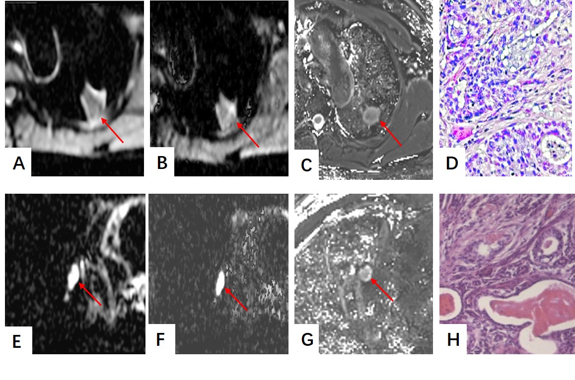
A-D A adenocarcinoma in the left upper lobe (A) An axial ADC mapping. (B) D map. (C) T2 map. (D) Hematoxylin-eosin staining confirms
the nodule which is associated with nuclear polymorphism, higher cellularity and nuclear-to-cytoplasmic ratios which make ADC value, D value, and T2 value
down.E-H A ball pneumonia in the right middle lobe. (E) An axial ADC mapping. (F) D map. (G) T2 map. (H) Hematoxylin-eosin staining confirms
the nodule was characterized by more extracellular fluid spec and tissue fluid
which would make ADC value, D value and T2 value rise.