James A Eaden1,2, Guilhem J Collier1, Ho-Fung Chan1, Nicholas D Weatherley1,2, Graham Norquay1, Smitha Rajaram3, Andy Swift1,3, Ronald A Karwoski4, Brian Bartholmai4, Stephen M Bianchi2, and Jim M Wild1,5
1POLARIS, MRI unit, Department of Infection Immunity and Cardiovascular Disease, University of Sheffield, Sheffield, United Kingdom, 2Academic Directorate of Respiratory Medicine, Sheffield Teaching Hospitals NHS Foundation Trust, Sheffield, United Kingdom, 3Sheffield Teaching Hospitals NHS Foundation Trust, Sheffield, United Kingdom, 4Mayo Clinic, Rochester, MN, United States, 5Insigneo institute, University of Sheffield, Sheffield, United Kingdom
1POLARIS, MRI unit, Department of Infection Immunity and Cardiovascular Disease, University of Sheffield, Sheffield, United Kingdom, 2Academic Directorate of Respiratory Medicine, Sheffield Teaching Hospitals NHS Foundation Trust, Sheffield, United Kingdom, 3Sheffield Teaching Hospitals NHS Foundation Trust, Sheffield, United Kingdom, 4Mayo Clinic, Rochester, MN, United States, 5Insigneo institute, University of Sheffield, Sheffield, United Kingdom
Several significant correlations were found between dissolved 129Xe MRI (RBC:TP, TP:Gas and RBC:Gas) and CALIPER
CT variables, both globally and regionally in IPF patients. There were no
significant correlations between 129Xe DW-MRI and CALIPER
variables.
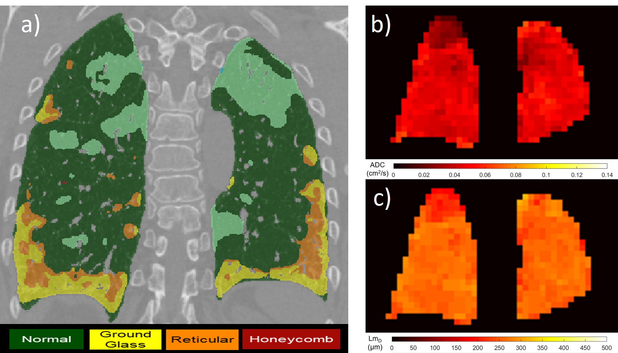
Figure 1. Examples of CALIPER CT (a), 129Xe
ADC (b) and 129Xe LmD (c) images.
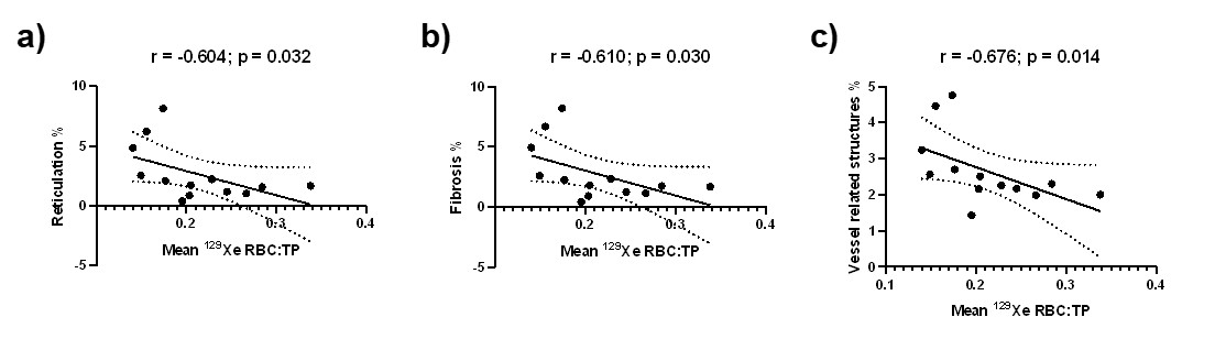
Figure 2. Correlation between global 129Xe RBC:TP and CALIPER reticulation % (a), fibrosis % (b) and vessel related
structures % (c) (n=13).