Laura Saunders1, Dave Capener1, David G Kiely1,2, Andy J Swift1, and Jim M Wild1
1Infection Immunity and Cardiovascular Disease, University of Sheffield, Sheffield, United Kingdom, 2Sheffield Pulmonary Vascular Disease Unit, Sheffield, United Kingdom
1Infection Immunity and Cardiovascular Disease, University of Sheffield, Sheffield, United Kingdom, 2Sheffield Pulmonary Vascular Disease Unit, Sheffield, United Kingdom
Lung T1 was significantly lower in patients with emphysema seen on
3D CT than in patients with fibrosis, centrilobular ground glass or no lung
disease seen on CT. Lung T1 was more sensitive in
differentiating lung pathology seen on CT when calculated using median T1 excluding lung vessels.
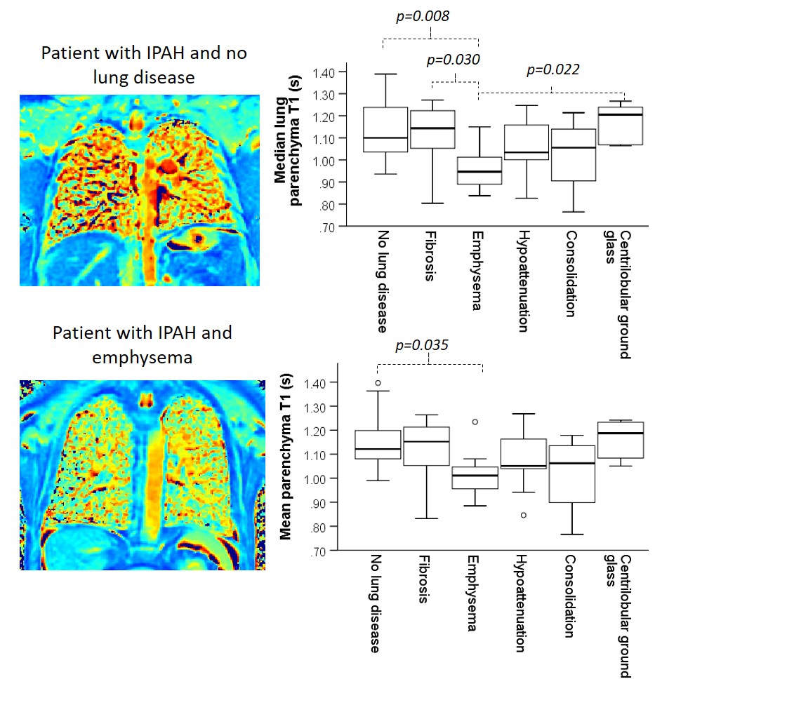
Figure 3: Two example T1 maps of patients with
IPAH with no lung disease seen on CT, and with emphysema seen on CT (left) and two
box plots of mean and median lung T1 in patients with PH (right).
There are significant differences in
median parenchyma T1 between patients with emphysema and patients
with fibrosis and centrilobular ground glass that are not seen when using mean
T1 as an average metric.
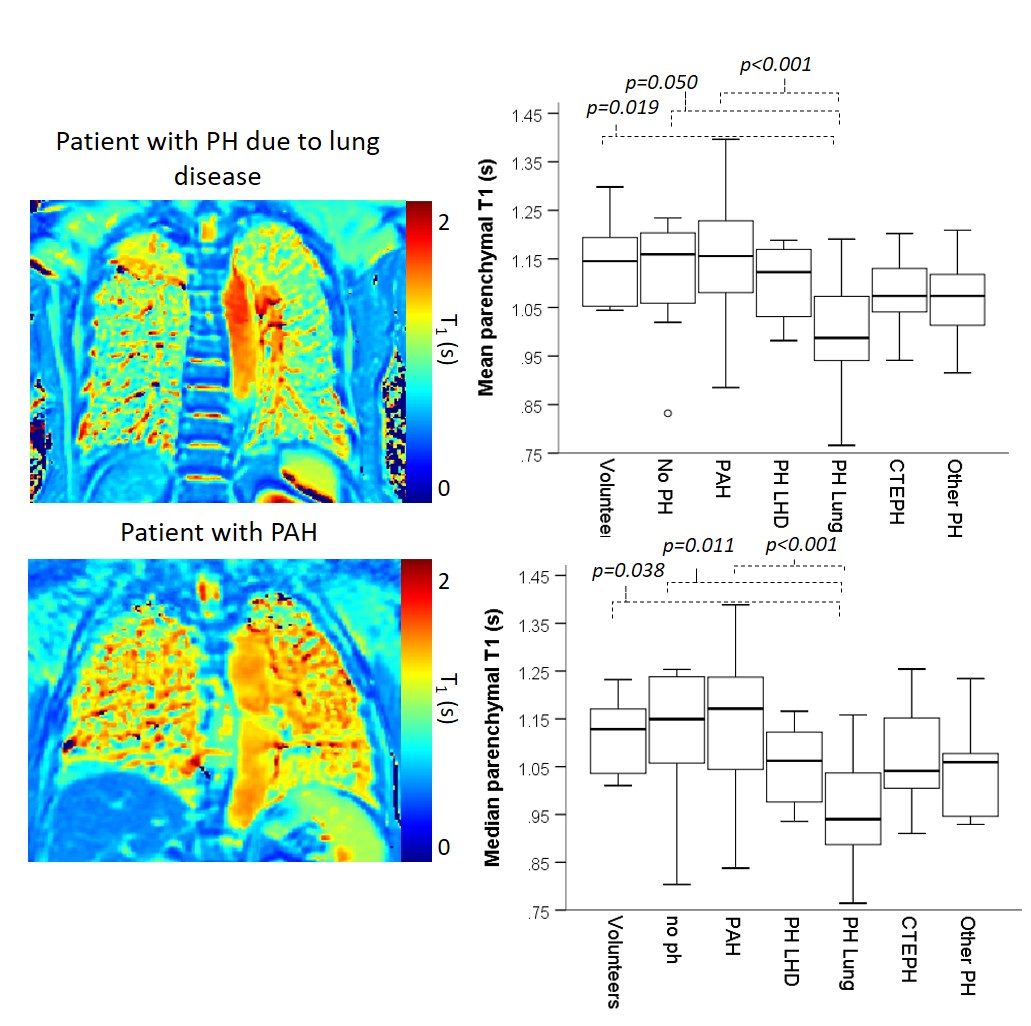
Figure 2: Example T1 maps in
a patient with PH due to lung disease and pulmonary arterial hypertension. Box plots
of mean and median lung T1 in patients with PH and control groups
both show significantly lower lung parenchyma T1 in patients with PH
due to lung disease, than patients without PH, patients with PAH or healthy volunteers.