El-Sayed H Ibrahim1, Abdul Parchur1, Brian Fish1, Meetha Medhora1, and Amit Joshi1
1Medical College of Wisconsin, Milwaukee, WI, United States
1Medical College of Wisconsin, Milwaukee, WI, United States
Ultrashort echo-time
dynamic contrast enhancement MRI has the potential for in vivo quantification
of irradiation induced vascular perfusion and permeability early changes in
multiple organs in the same subject.
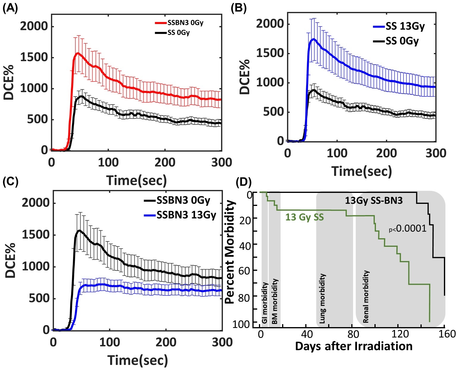
Figure 1. (a) Ultrashort echo-time dynamic contrast
enhancement MRI image and signal enhancement evolution in the lung before (baseline)
and after gadolinium injection in (A) SS and SSBN3 rats (control- 0 Gy),(B) SS
rats radiated at 0 (n = 4) and 13 Gy (n = 5), and (C) SSBN3 rats radiated at 0 (n
= 4) and 13 Gy (n = 4). Error bars represent mean ± SEM. (D) Survival curve Leg-out
PBI with supportive care in SS and SS-BN3 rats.
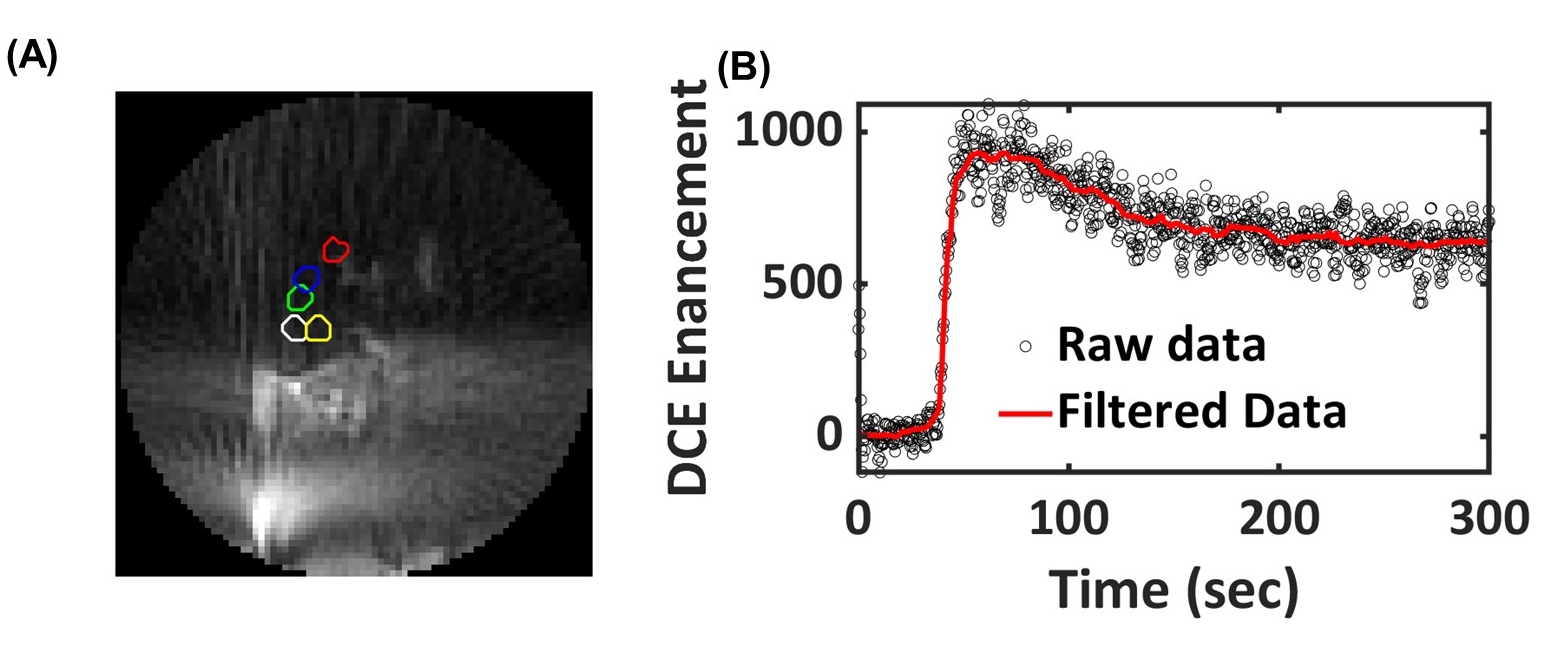
Figure 2. (A) A typical
image of ultrashort echo-time dynamic contrast enhancement MRI image in the
lung (five ROIs in the lung used to understand the perfusion in the lung) and
(B) kinetic DCE raw data (without motion correction – black circles) and
filtered data for respiratory motion correction (red line).