Laurence H. Jackson1, Joanna Bell2, Giulia Benedetti2, Sze Mun Mak2, Rachael Franklin3, Rebecca Preston2, Andrea Bille4, John Spence2, and Geoff Charles-Edwards1
1Medical Physics, Guy's and St Thomas' NHS Foundation Trust, London, United Kingdom, 2Department of Radiology, Guy's and St Thomas' NHS Foundation Trust, London, United Kingdom, 3Medical Physics, King's College Hospital NHS Foundation Trust, London, United Kingdom, 4Department of Thoracic Surgery, Guy's and St Thomas' NHS Foundation Trust, London, United Kingdom
1Medical Physics, Guy's and St Thomas' NHS Foundation Trust, London, United Kingdom, 2Department of Radiology, Guy's and St Thomas' NHS Foundation Trust, London, United Kingdom, 3Medical Physics, King's College Hospital NHS Foundation Trust, London, United Kingdom, 4Department of Thoracic Surgery, Guy's and St Thomas' NHS Foundation Trust, London, United Kingdom
Here we present an MRI method for the pre and postoperative assessment of chest wall and diaphragmatic motion which provides valuable dynamic information to aid radiological assessment in these highly variable patients.

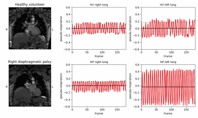
Figure 4. Comparison of right and left lung xt-PCA tidal breathing signal in a healthy volunteer (top row) and in a patient with right diaphragmatic palsy (DP) (bottom row). The dysfunction of the right lung in the patient manifests as a disproportionate diaphragmatic excursion with a compensatory larger signal in the left lung.

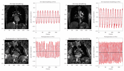
Figure 3. Results of xt-PCA diaphragm motion extraction in a healthy volunteer (top row) and a postsurgical patient following robotic plication and chest wall reconstruction (bottom row). A comparison of tidal breathing and maximum inspiration and expiration shows a number of potentially useful features for diagnosis, including amplitude, frequency and regularity. Both figures are animated to the same time scale and included on each figure is the mean signal (black) and ±1σ standard deviation (grey).
