Bilal A Tahir1,2, Laurie J Smith1, Joshua R Astley1,2, Michael Walker1, Alberto M Biancardi1, Guilhem J Collier1, Paul J Hughes1, Helen Marshall1, and Jim M Wild1
1POLARIS, University of Sheffield, Sheffield, United Kingdom, 2Oncology and Metabolism, University of Sheffield, Sheffield, United Kingdom
1POLARIS, University of Sheffield, Sheffield, United Kingdom, 2Oncology and Metabolism, University of Sheffield, Sheffield, United Kingdom
This study compares surrogates of
regional ventilation, derived from inspiratory and expiratory 3D proton MRI
with hyperpolarized gas MRI, pulmonary functions tests and multiple-breath
washout, in patients with a broad range of cystic fibrosis disease severity and
age.
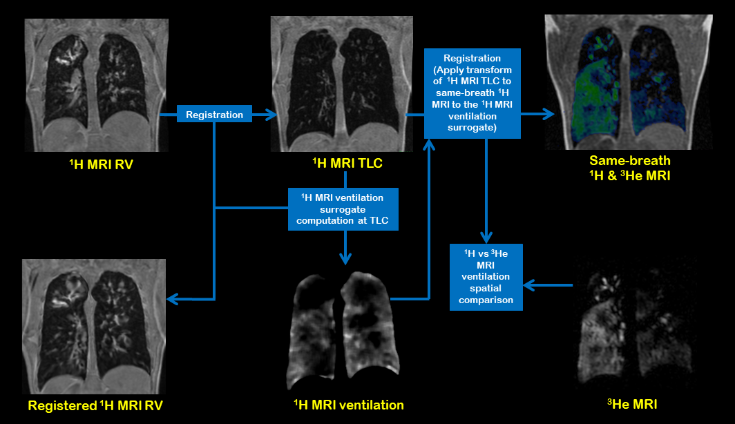
Figure 1. Workflow of
spatial comparison method of 1H and 3He MRI ventilation.
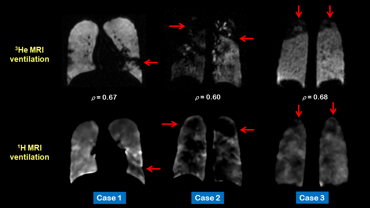
Figure 2. Corresponding coronal slices for three
patients of 3He (top row)) and 1H (bottom row) MRI
ventilation after registration. The red arrows depict defects and regions that
are visually similar on both scans. Voxel-wise Spearman's correlation
coefficients (ρ) are provided for each case.