Mary A. Neal1,2, Benjamin J. Pippard1,2, Kieren G. Hollingsworth1,2, A. John Simpson2, and Peter E. Thelwall1,2
1Newcastle Magnetic Resonance Centre, Newcastle University, Newcastle upon Tyne, United Kingdom, 2Translational and Clinical Research Institute, Newcastle University, Newcastle upon Tyne, United Kingdom
1Newcastle Magnetic Resonance Centre, Newcastle University, Newcastle upon Tyne, United Kingdom, 2Translational and Clinical Research Institute, Newcastle University, Newcastle upon Tyne, United Kingdom
Dynamic 19F-MRI of inhaled perfluoropropane for regional assessment of gas wash-in and wash-out rates has been demonstrated in patients with COPD using compressed sensing for scan acceleration. A strong correlation with spirometric FEV1 was measured.
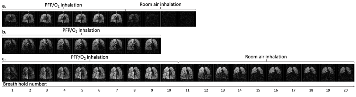
Figure 1: Multi-breath wash-in and wash-out of inhaled PFP/O2. A central coronal slice from a 3D 19F-MR image acquired during each breath hold is displayed. Images were acquired on alternate inhalations (ie. after 1, 3, 5, etc. inhalations of PFP/O2). PFP/O2 wash-in is visible over the initial breaths. The inhalation rig valve was then switched to deliver room air for visualisation of PFP/O2 gas wash-out. 1a. PFP/O2 wash-in and wash-out in a healthy volunteer. 1b. PFP/O2 wash-in in P1 (nb. wash-out images were not acquired in this participant). 1c. PFP/O2 wash-in and wash-out in P2.
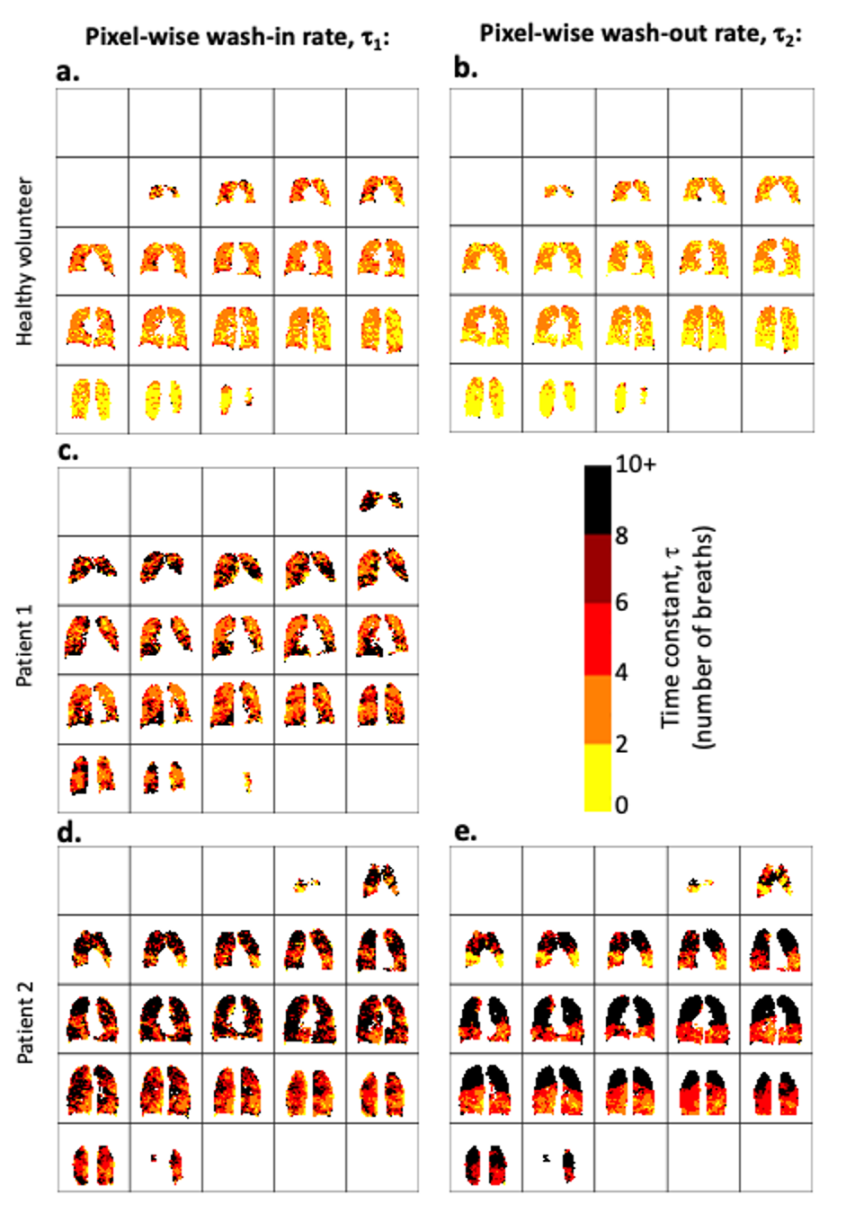
Figure 3: 3D voxel-wise exponential time constant estimation, where τ1 represents the number of wash-in respiratory cycles required to increase the 19F-MRI signal to 1 - 1/e (≈63.2%) of its calculated maximum value. τ2 represents the number of respiratory cycles required to reduce the 19F-MRI signal to 1/e (≈36.8%) of its calculated maximum value. Maps for wash-in τ1(3a., 3c., and 3d.) and wash-out τ2 (3b. and 3e.) are shown.