Seokwon Lee1, Jinil Park2, Hyonha Kim1, Ho Yun Lee3, and Jang-Yeon Park1,4
1Department of Biomedical Engineering, Sungkyunkwan University, Suwon, Korea, Republic of, 2Biomedical Institute for Convergence at SKKU, Sungkyunkwan University, Suwon, Korea, Republic of, 3Department of Radiology and Center for Imaging Science, Samsung Medical Center, Seoul, Korea, Republic of, 4Department of Intelligent Precision Healthcare Convergence, Sungkyunkwan University, Suwon, Korea, Republic of
1Department of Biomedical Engineering, Sungkyunkwan University, Suwon, Korea, Republic of, 2Biomedical Institute for Convergence at SKKU, Sungkyunkwan University, Suwon, Korea, Republic of, 3Department of Radiology and Center for Imaging Science, Samsung Medical Center, Seoul, Korea, Republic of, 4Department of Intelligent Precision Healthcare Convergence, Sungkyunkwan University, Suwon, Korea, Republic of
This
study shows functional information such as ventilation and ventilation flow maps
including the histogram can be used to diagnose COPD phenotypes and disease
progression, together with structural information of UTE-MRI.
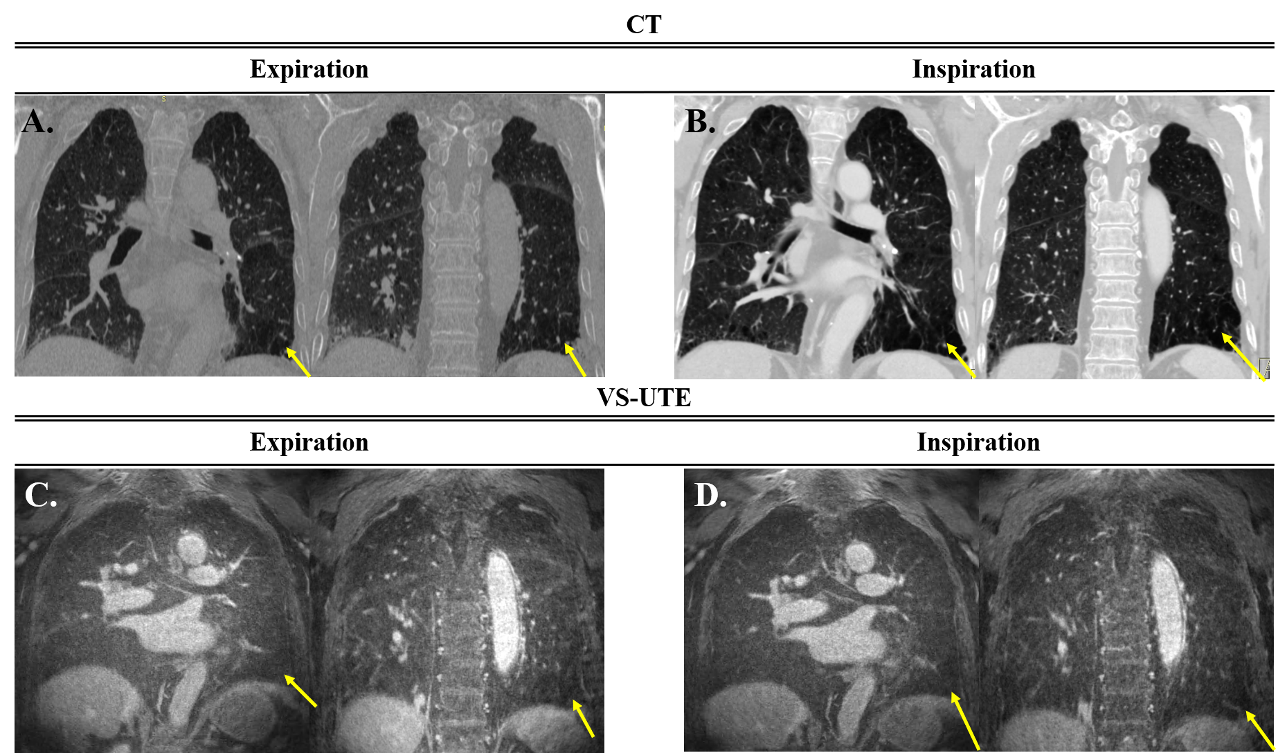
Figure 1.(A) CT images at expiration
state, (B) CT images at inspiration state, (C) MR images at expiration state, (D)
MR images at inspiration state. Yellow arrows indicate emphysema lesion. In
VS-UTE, although there is a slight decrease in signal, it is not easy to
accurately diagnose the lesion.
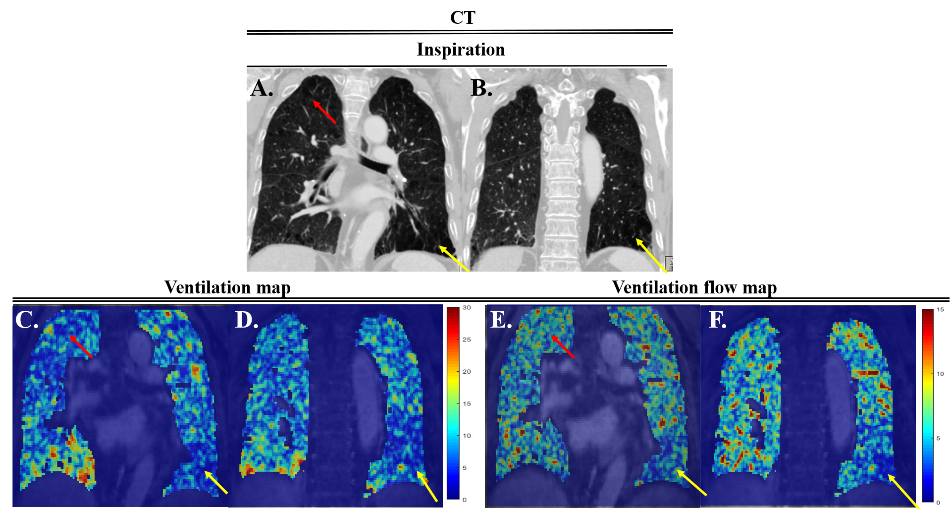
Figure
2. (A,B)
CT image at inspiration state, (C,D) Ventilation map each slide using VS-UTE,
(E,F) Ventilation flow map each slide using VS-UTE. Yellow arrows indicate
emphysema lesion. Figure 2 show the left lower lobe had high correlation with
CT and MRI ventilation map.