Fabien J Bertin1, Guilhem J Collier2, Paul JC Hughes2, Laurie Smith2, James Aeden2, Helen Marshall2, Jim M Wild2,3, and Alberto M Biancardi2
1Télécom SudParis, Paris, France, 2POLARIS, Department of Infection, Immunity and Cardiovascular Disease, The University of Sheffield, Sheffield, United Kingdom, 3Insigneo Institute for in silico Medicine, The University of Sheffield, Sheffield, United Kingdom
1Télécom SudParis, Paris, France, 2POLARIS, Department of Infection, Immunity and Cardiovascular Disease, The University of Sheffield, Sheffield, United Kingdom, 3Insigneo Institute for in silico Medicine, The University of Sheffield, Sheffield, United Kingdom
A specific tailoring
of a well-known Deep-Learning approach was developed to automate the identification
of airways and ventilated lungs in pulmonary hyperpolarised-gas MR images. A
good performance was achieved, capable of discriminating between ambiguous
features.
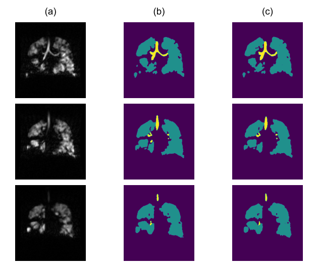
Figure 1: An example of the
segmentation performance. Three consecutive slices of a 3D image are shown in
column (a) together with the manual ground truth in (b) and the DL output in
(c), with parenchyma in green and airways in yellow. The example shows the
quality of the predictions and the challenges the system has to face, where a
small portion of one airway in the mid slice was excluded from both labels.
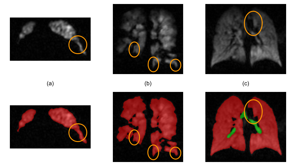
Figure 2: Examples of HP gas
images where some ambiguous features, circled in orange, are present. These
features resemble airway segments and, potentially, could be misclassified. In the upper row the HP gas images are shown,
in the lower row the DL output is displayed, which shows that the features were
correctly identified as belonging to the lung ventilated region.