Sarah H. Needleman1, Jamie R. McClelland2, Björn Eiben2, and Geoff J. M. Parker1,3
1Centre for Medical Image Computing, Quantitative Imaging Group, Department of Medical Physics and Biomedical Engineering, University College London, London, United Kingdom, 2Centre for Medical Image Computing, Radiotherapy Image Computing Group, Department of Medical Physics and Biomedical Engineering, University College London, London, United Kingdom, 3Bioxydyn Limited, Manchester, United Kingdom
1Centre for Medical Image Computing, Quantitative Imaging Group, Department of Medical Physics and Biomedical Engineering, University College London, London, United Kingdom, 2Centre for Medical Image Computing, Radiotherapy Image Computing Group, Department of Medical Physics and Biomedical Engineering, University College London, London, United Kingdom, 3Bioxydyn Limited, Manchester, United Kingdom
The simulated dynamic oxygen-enhanced MRI series produced by
the framework was realistic, displayed respiratory motion, and quantitative
measures describing hyperoxia-induced contrast enhancement agreed with
experimental literature.
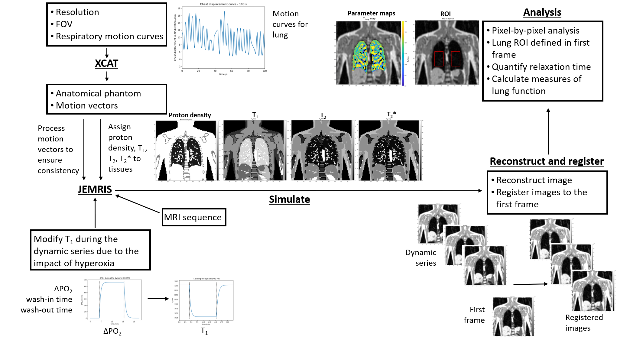
Figure 1: Representation of the pipeline of processes which
formed the framework which was applied to model dynamic OE-MRI scans.
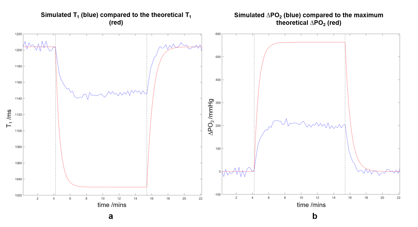
Figure 3: Comparison of predicted (calculated using a value
for the maximum theoretical ΔPO2) T1(t) (a)
and ΔPO2(t)
(b) to values extracted from the dynamic series. The extracted T1
and ΔPO2
values change by less than predicted. Plateau T1,oxy = (1147 ±
3) ms; ΔPO2
= (201 ± 9) mmHg. Vertical lines indicate a switch from the simulated
subject breathing air to 100% O2 (4.2 minutes) or 100% O2
to air (15.4 minutes).