Brandon Zanette1, Yonni Friedlander1,2, Marcus J Couch3,4,5, and Giles Santyr1,2
1Translational Medicine, The Hospital for Sick Children, Toronto, ON, Canada, 2Department of Medical Biophysics, University of Toronto, Toronto, ON, Canada, 3Siemens Healthcare Limited, Montreal, QC, Canada, 4McConnell Brain Imaging Centre, Montreal Neurological Institute, Montreal, QC, Canada, 5Department of Neurology and Neurosurgery, McGill University, Montreal, QC, Canada
1Translational Medicine, The Hospital for Sick Children, Toronto, ON, Canada, 2Department of Medical Biophysics, University of Toronto, Toronto, ON, Canada, 3Siemens Healthcare Limited, Montreal, QC, Canada, 4McConnell Brain Imaging Centre, Montreal Neurological Institute, Montreal, QC, Canada, 5Department of Neurology and Neurosurgery, McGill University, Montreal, QC, Canada
A spiral imaging
sequence was developed and tested in a 19F phantom to improve SNR and reduce acquisition
times. A 44% improvement in SNR was observed in 1/3 of the acquisition time
compared to a typical GRE sequence, indicating potential to improve in-vivo 19F
gas MRI image quality in the future.

Figure
1:
Representative 19F gas images acquired with (a) 2D-GRE and (b)
3D-SoS. The yellow squares approximate the 9×9 ROI and locations in the phantom
and background used to measure mean signal and background noise for SNR
measurements.
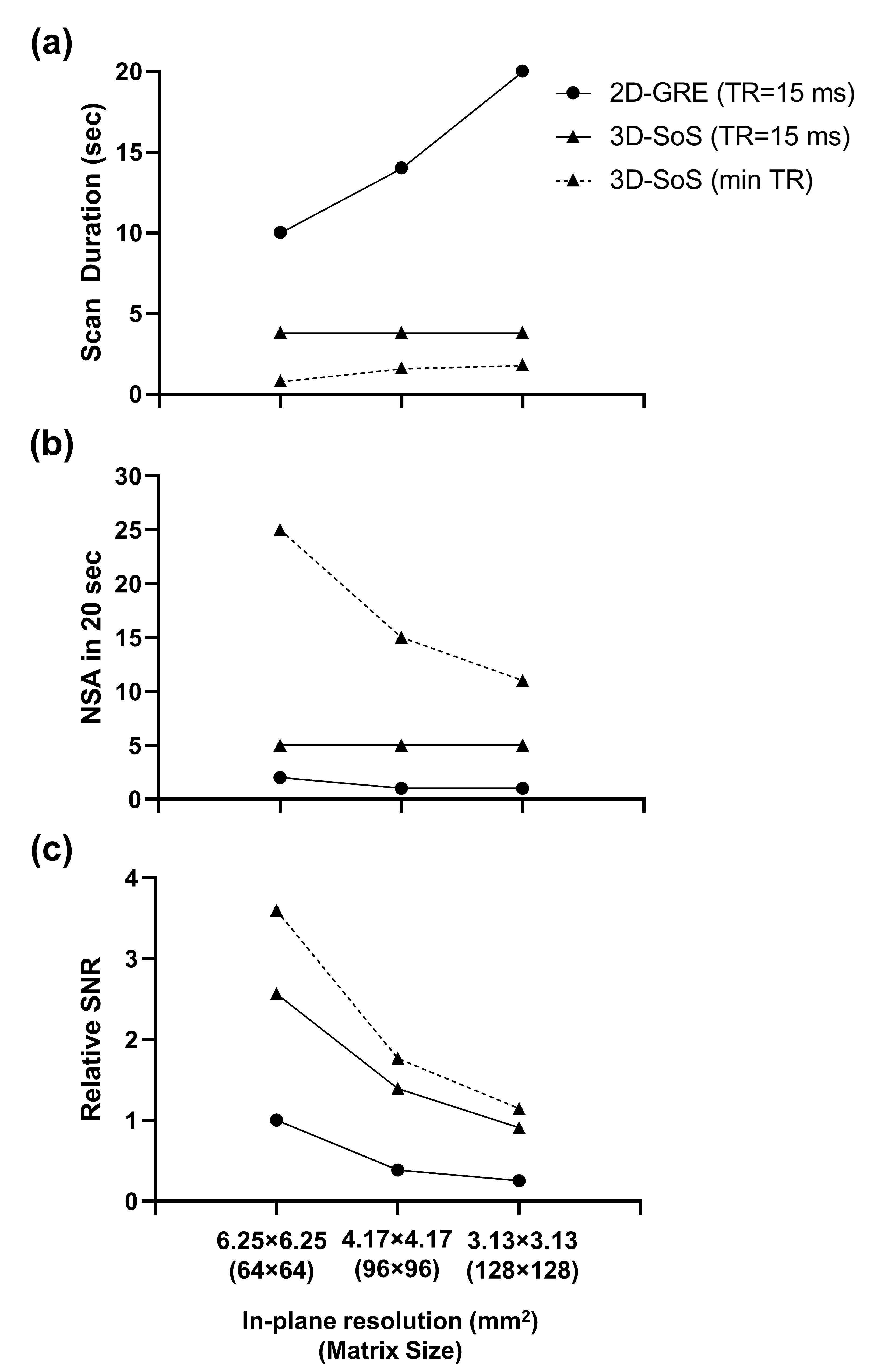
Figure 2: (a)
Simulated durations for three clinically relevant in-plane resolutions for
2D-GRE and 3D-SoS at a constant TR of 15 ms (10 slices). All other parameters
are as stated in Table 1 for each sequence. A simulation with the minimum TR
for 3D-SoS is also shown. (b) Achievable number of signal averages (NSA)
in an assumed 20 second breath-hold, rounded to the nearest integer using scan
durations in (a). (c) Relative SNR accounting for changes in spatial
resolution, NSA in 20 sec, and difference in TE. All SNRs normalized to the 64×64
2D-GRE.