Efe Ilicak1, Jascha Zapp1, Safa Ozdemir1, Lothar R. Schad1, and Frank G. Zöllner1,2
1Computer Assisted Clinical Medicine, Medical Faculty Mannheim, Heidelberg University, Mannheim, Germany, 2Mannheim Institute for Intelligent Systems in Medicine, Medical Faculty Mannheim, Heidelberg University, Mannheim, Germany
1Computer Assisted Clinical Medicine, Medical Faculty Mannheim, Heidelberg University, Mannheim, Germany, 2Mannheim Institute for Intelligent Systems in Medicine, Medical Faculty Mannheim, Heidelberg University, Mannheim, Germany
Fourier
Decomposition MRI uses bSSFP sequence for assessing pulmonary functions.
However, it suffers from banding artefacts. We propose phase-cycled acquisitions
for improved robustness. In vivo results from 1.5 T and 3 T scanners are
provided to demonstrate the proposed method.
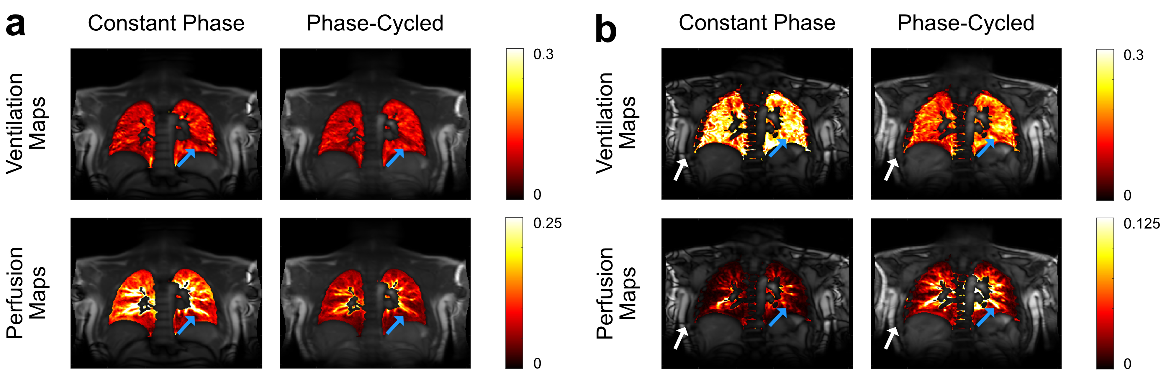
Figure
3: Combined functional maps overlaid on
a cross-section shown for 1.5 T (a) and 3 T (b). At 1.5T, both phase-cycled maps
provide similar contrast and prominent structures (blue arrows) compared to
constant phase maps while phase-cycled perfusion map suffers from overall lower
values due to averaging. At 3T, both phase-cycled
maps show similar contrast and prominent structures (blue arrows) compared to
1.5T. However, the constant phase is less able to reproduce these structures since
it is more prone to field
inhomogeneity artefacts
(white arrows).
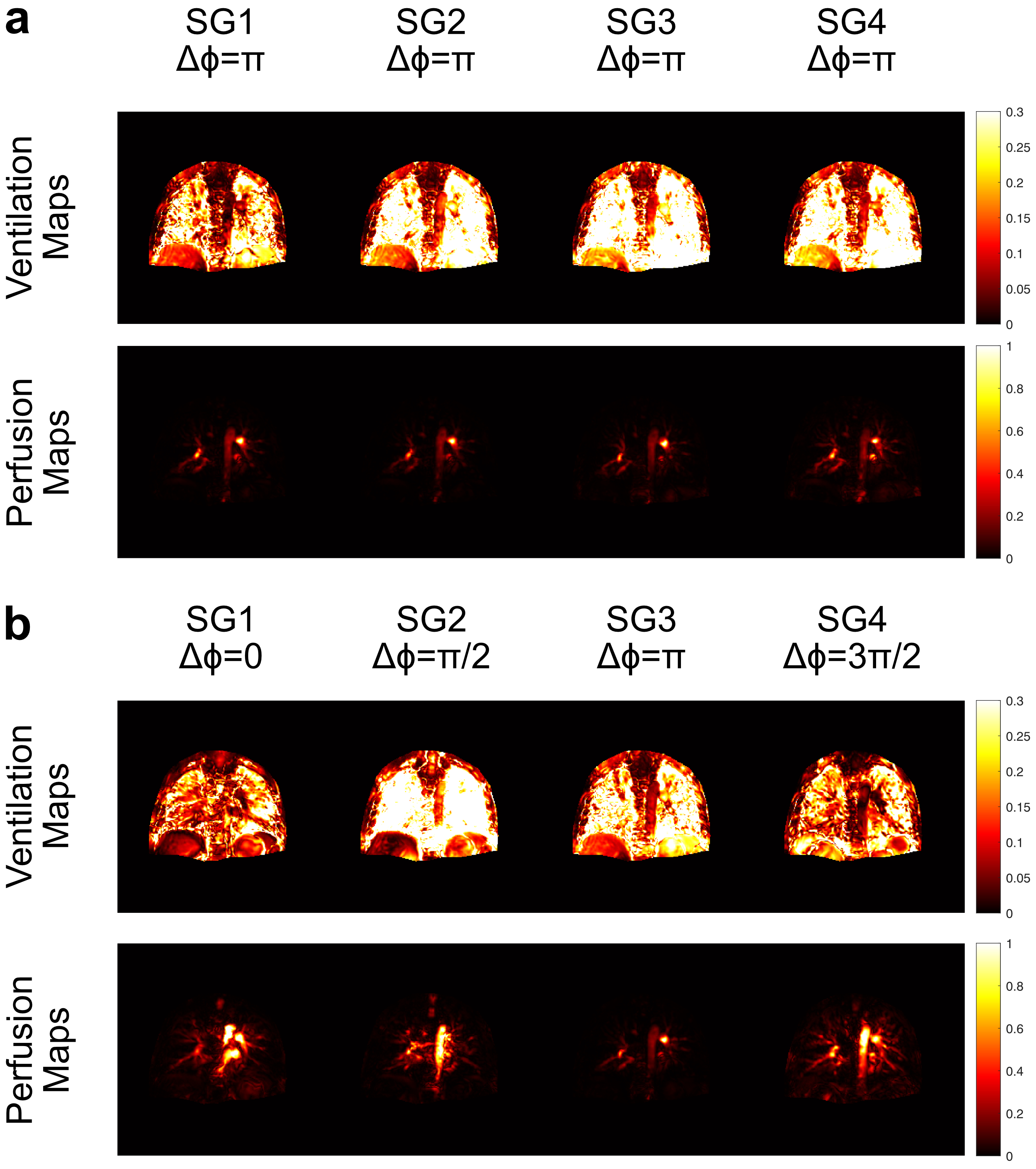
Figure 2:
Ventilation and perfusion maps of subgroups at 3 T for constant phase (a) and
phase-cycled (b) bSSFP acquisitions. As expected, constant phase acquisition
generates similar functional maps throughout the experiment whereas
phase-cycled acquisition is able to generate functional maps with different
information by changing the RF phase. At 3 T, the perfusion maps with
conventional $$$\Delta\phi = \pi$$$ acquisition suffer
from field inhomogeneity and are less comprehensive compared to phase-cycled
acquisitions.