Grzegorz Bauman1,2 and Oliver Bieri1,2
1Department of Radiology, Division of Radiological Physics, University of Basel Hospital, Basel, Switzerland, 2Department of Biomedical Engineering, University of Basel, Allschwil, Switzerland
1Department of Radiology, Division of Radiological Physics, University of Basel Hospital, Basel, Switzerland, 2Department of Biomedical Engineering, University of Basel, Allschwil, Switzerland
We demonstrate the feasiblity of free-breathing respiratory self-gated thoracic MRI with balanced steady-state free precession half-radial dual-echo imaging technique (bSTAR) in human subjects.
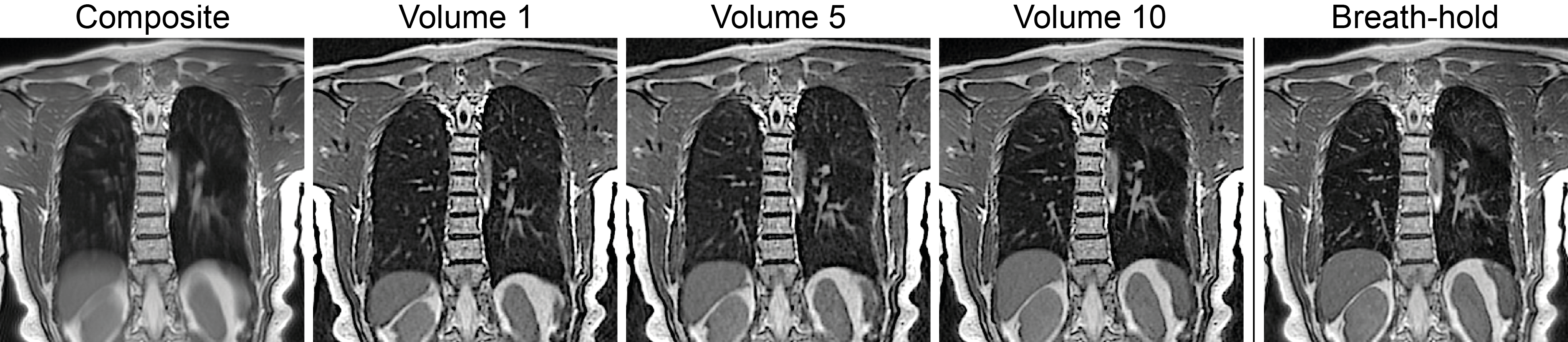
Figure 3. Illustrative
coronal FB bSTAR image reconstructions in a healthy volunteer: using all
acquired data (no gating) - composite (a), data binned to end inspiration -
volume 1 (b), data binned to intermediate respiratory state - volume 5 (c) and data binned to end
expiration - volume 10 (d). For comparison breath-hold bSTAR acquisition was
performed (e).
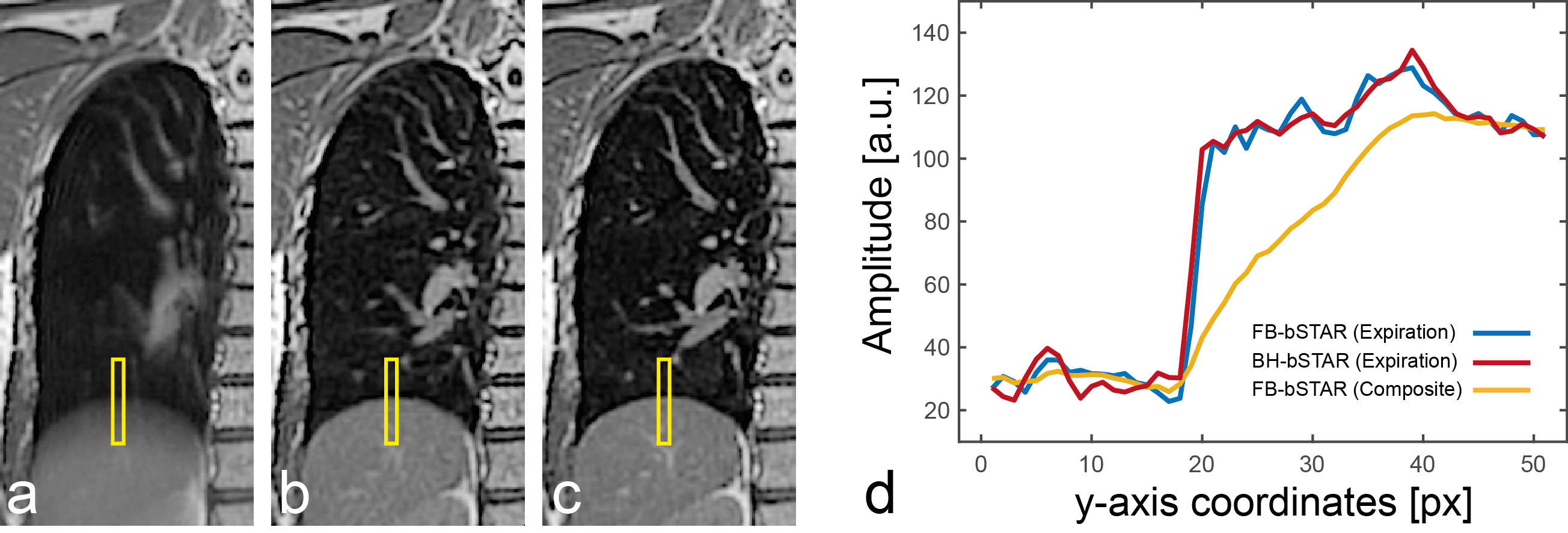
Figure 4. Comparison
between free-breathing bSTAR composite (a), free-breathing bSTAR expiratory
reconstruction (b), and expiratory breath-hold bSTAR image. Diagram (d) shows
signal amplitude profiles obtained in the region indicated by the yellow box
(signal amplitude was averaged along the x-axis).