Vadim Malis1, Won Bae1, Asako Yamamoto1, Yoshimori Kassai2, Andrew Yen1, Susan Hopkins1, Yoshiharu Ohno3, and Mitsue Miyazaki1
1Radiology, UC San Diego, San Diego, CA, United States, 2Canon Medical, Tochigi, Japan, 3Radiology, Fujita Health University, Toyoake, Japan
1Radiology, UC San Diego, San Diego, CA, United States, 2Canon Medical, Tochigi, Japan, 3Radiology, Fujita Health University, Toyoake, Japan
Free breathing lung MRI techniques were developed for measurement of T2*, inspiratory/expiratory lung volumes, specific ventilation and visualization of the pulmonary vasculature.
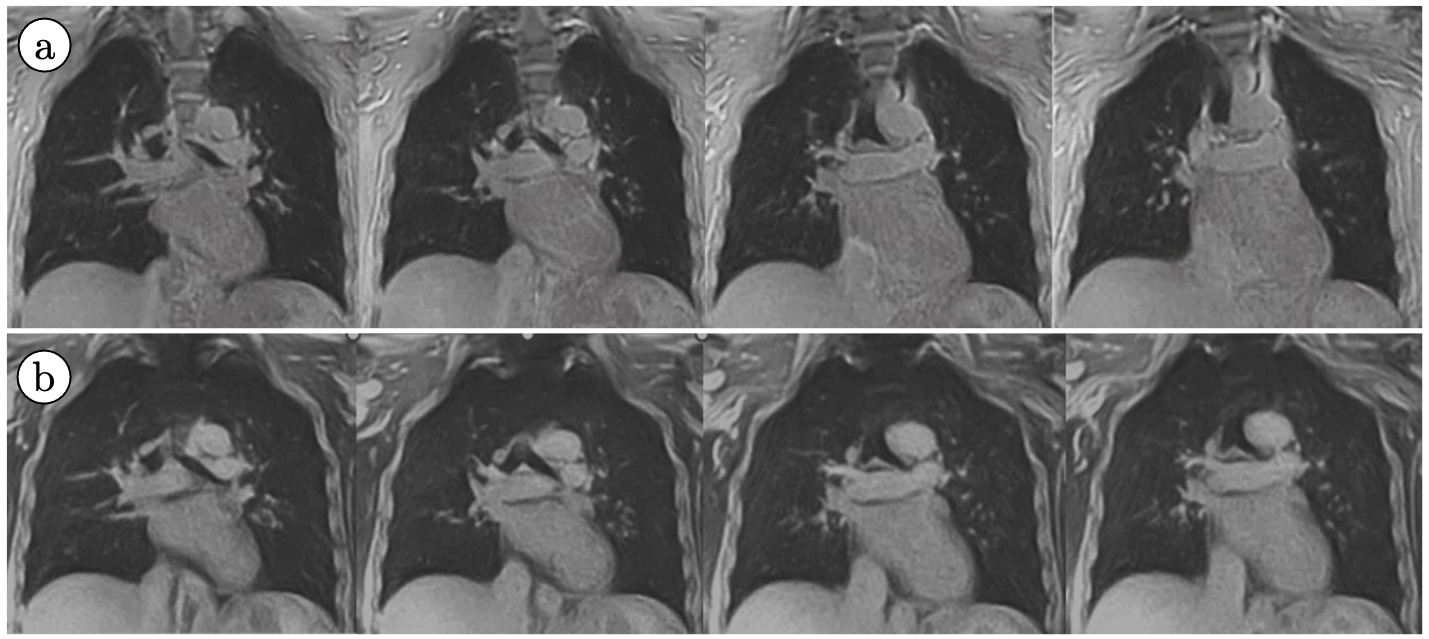
Figure 3: Free-breathing 3D UTE images of 4 consecutive slices acquired without fat suppression (a) and with fat suppression (b). Fat around the heart is suppressed in (b) with better visualization of pulmonary arteries and myocardium.
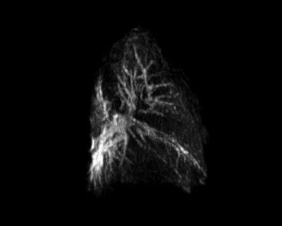
Figure 4: Maximum Intensity Projection of a segmented lung volume from fat-suppressed 3D UTE.