Ahsan Javed1, Rajiv Ramasawmy1, Pan Su2, Thomas Benkert3, Waqas Majeed2, and Adrienne E Campbell-Washburn1
1Cardiovascular Branch, Division of Intramural Research, National Heart, Lung, and Blood Institute, National Institutes of Health, Bethesda, MD, United States, 2Siemens Medical Solutions USA Inc., Malvern, PA, United States, 3Siemens Healthcare GmbH, Erlangen, Germany
1Cardiovascular Branch, Division of Intramural Research, National Heart, Lung, and Blood Institute, National Institutes of Health, Bethesda, MD, United States, 2Siemens Medical Solutions USA Inc., Malvern, PA, United States, 3Siemens Healthcare GmbH, Erlangen, Germany
Free-breathing self-gated stack-of-spirals UTE can improve the sensitivity and repeatability of oxygen-enhanced lung imaging on high performance low-field systems by improving the signal-to-noise ratio, and consistency of respiratory position between scans.
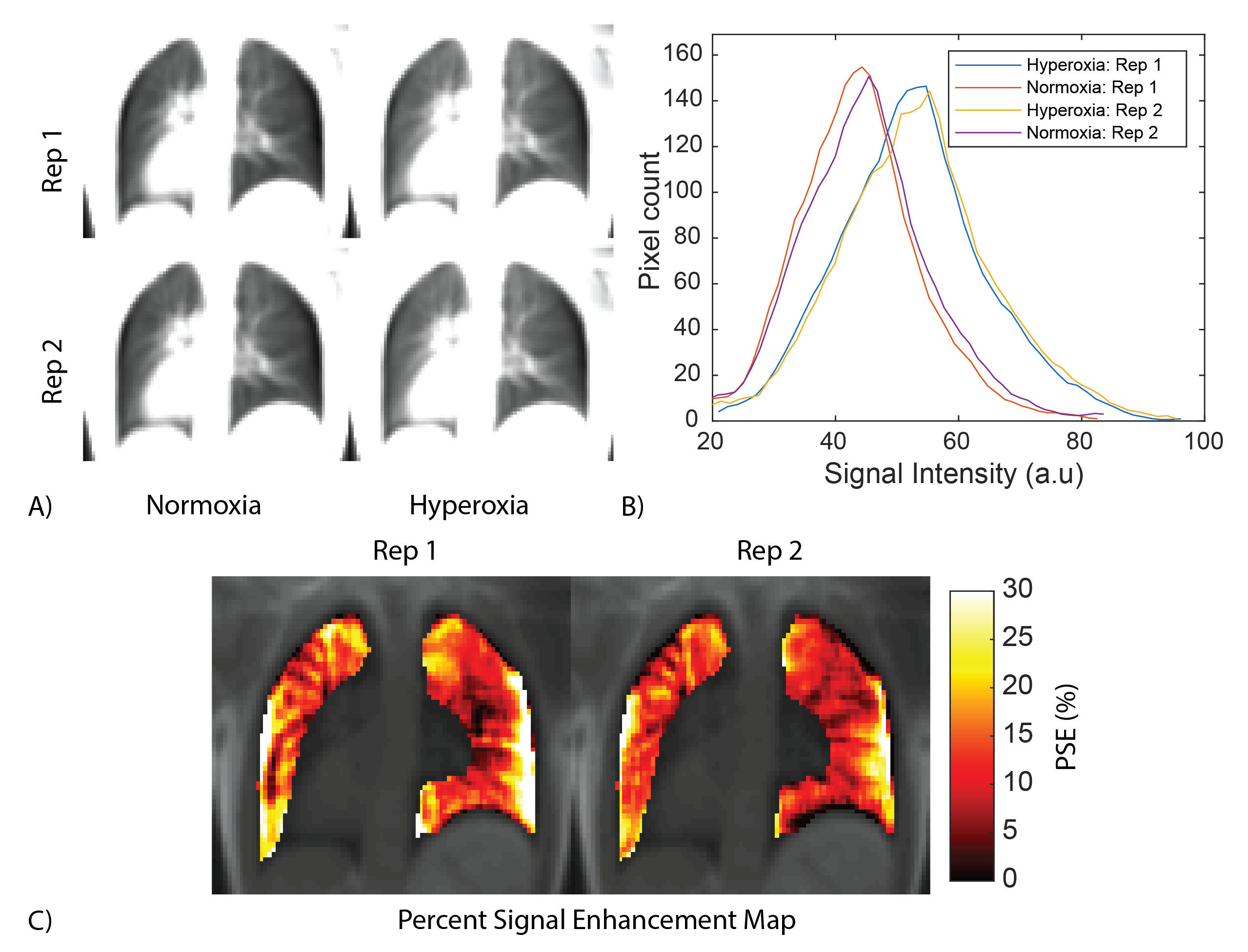
Figure 2: Signal enhancement in FB T1 weighted 3D stack of spiral UTE images. A) T1w UTE images at normoxia, and hyperoxia for two repetitions B) Histogram of signal intensity for normoxia and hyperoxia images within the lung for two repetitions shows excellent repeatability. C) PSE maps for the two repitions estimated from images in A) using Eq 1.
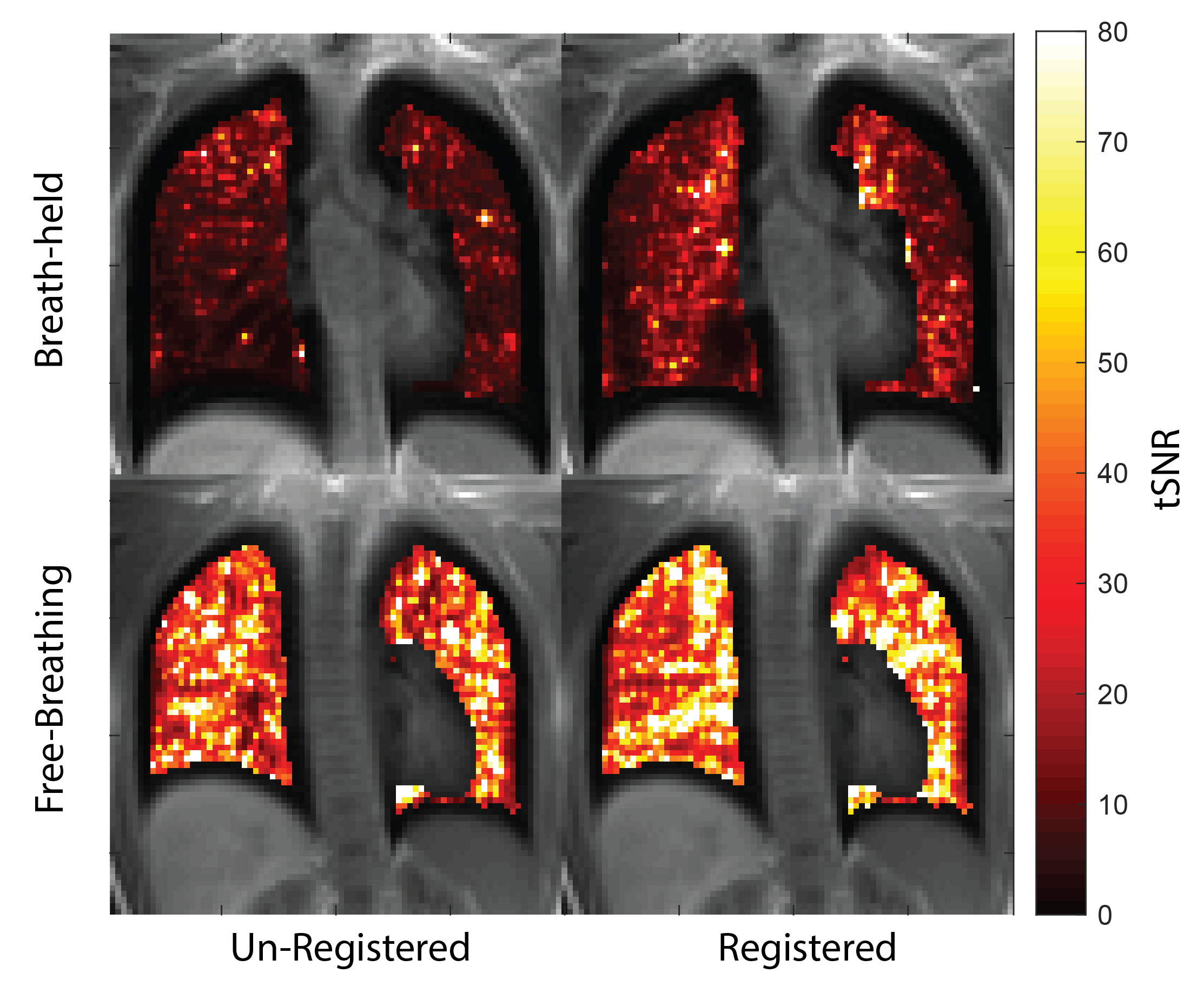
Figure 4: Representative temporal SNR map from subject 2. Images reconstructed from (top) BH data and (bottom) FB data. (left) Images before registration and (right) images after registration. tSNR is significantly higher for FB (un-registered: 23.8 ± 13.7, registered: 27.1+/-13.6) data compared to BH (un-registered: 9.6 ± 1.5, registered: 12.6 ± 2.6). Registration improves tSNR, as expected.