Fei Tan1, Xucheng Zhu2, and Peder E.Z. Larson1,3
1Bioengineering, UC Berkeley - UCSF, San Francisco, CA, United States, 2GE Healthcare, Menlo Park, CA, United States, 3Radiology and Biomedical Imaging, University of California, San Francisco, San Francisco, CA, United States
1Bioengineering, UC Berkeley - UCSF, San Francisco, CA, United States, 2GE Healthcare, Menlo Park, CA, United States, 3Radiology and Biomedical Imaging, University of California, San Francisco, San Francisco, CA, United States
The reproducibility study results are shown by regional ventilation maps, split violin plots, within-subject coefficient of variation, Bland-Altman and linear regression plots. The Jacobian determinant based regional ventilation and the three registration methods are reproducible.
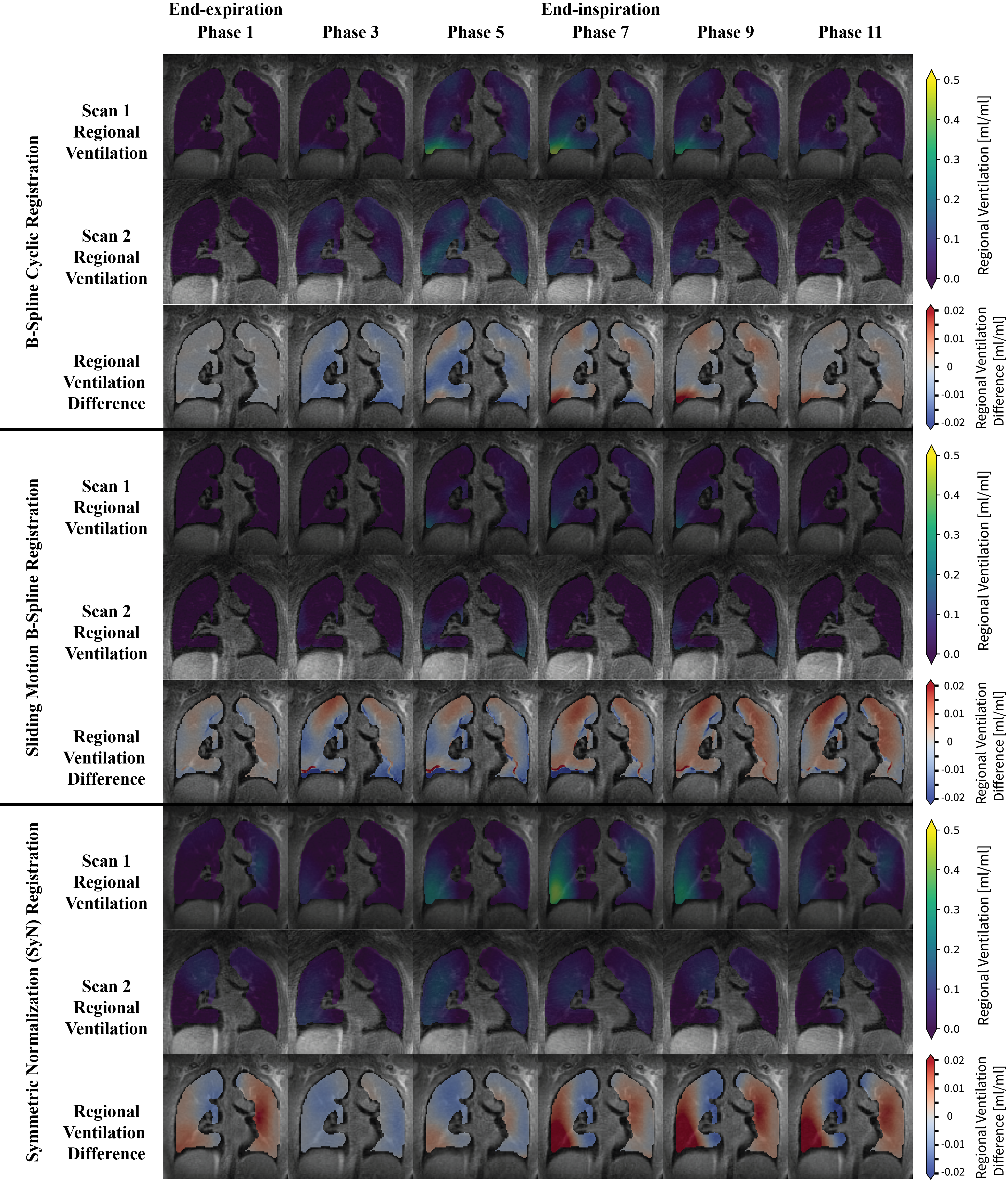
Figure 2. Representative Regional Ventilation Map of Two Scans and Their Difference Using Three Registration Methods. Within each method, the first row and second row are from the first and second scan respectively while the third row is the difference of ventilation maps between the two after a simple registration. Regional ventilation of 0.1 corresponds to 10% of volume expansion with respect to the end-expiration state. For conciseness, every other respiratory phase is shown.
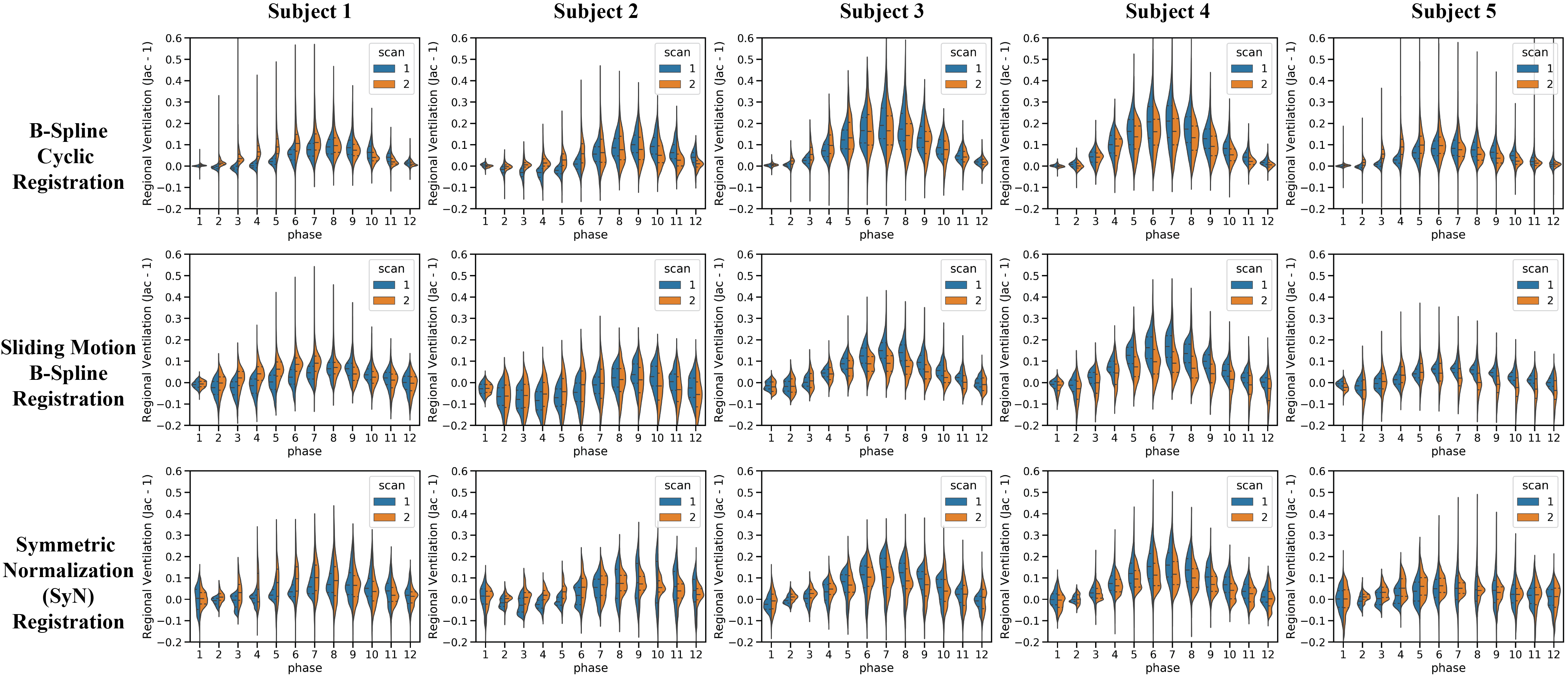
Figure 3. Split Violin Plot of the Regional Ventilation Distribution of All Subjects and Registration Methods. The discreet horizontal axis is the respiratory phases starting from the end-expiration phase, while the vertical axis is the regional ventilation. The two colors represent the 1st and 2nd scan respectively, and the shaded areas depict the regional ventilation distribution. The dotted lines are the quartiles of the distributions.