Anke Balasch1, Patrick Metze1, Kilian Stumpf1, Meinrad Beer2, Wolfgang Rottbauer1, and Volker Rasche1
1Department of Internal Medicine II, Ulm University Medical Center, Ulm, Germany, Ulm, Germany, 2Department of Radiology, Ulm University Medical Centre, Ulm, Germany, Ulm, Germany
1Department of Internal Medicine II, Ulm University Medical Center, Ulm, Germany, Ulm, Germany, 2Department of Radiology, Ulm University Medical Centre, Ulm, Germany, Ulm, Germany
Free
breathing and breathhold tyGA SoS imaging allows for sensitive assessment of
the morphology and function of the lung. The unique sampling pattern yield
excellent artifact properties in case of residual motion or irregular respiration.
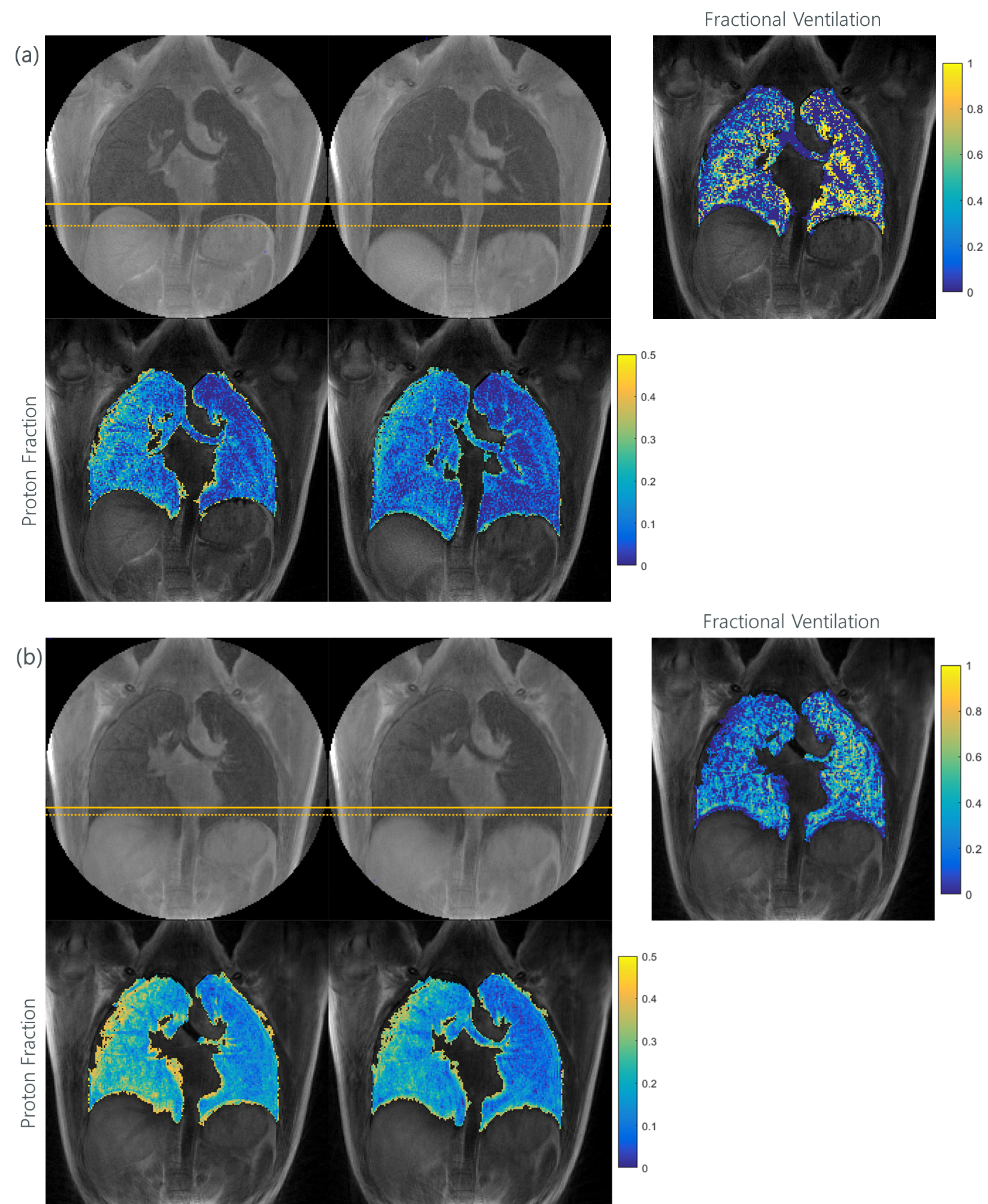
Figure 1: Example for BH (a) and SG (b) images in
end-expiration and end-inspiration. It can be seen, that the breathing
amplitude is bigger in the BH acquisition than in the SG. It is also visible,
the BH images are sharper than the SG images. For both techniques a FV-map
could be calculated. The proton fraction maps also shown. It is clearly
difference between EX and IN visible.
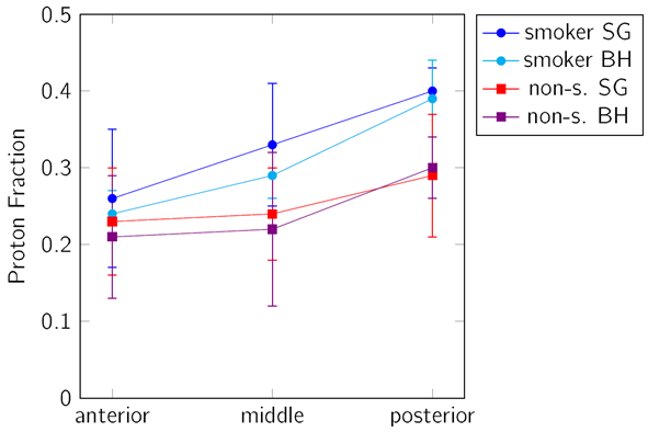
Figure 2: For smoker and non-smoker the proton fraction
values (the shown values are for EX) show a trend to increase from anterior to
posterior. The values for the smokers are significant higher than for the
non-smokers. The proton density in the BH images were a bit lower than in the
SG images.