Alexander German1, Angelika Mennecke1, Jan Martin2, Jannis Hanspach1, Andrzej Liebert1, Jürgen Herrler1, Tristan Anselm Kuder3, Manuel Schmidt1, Armin Nagel1, Michael Uder1, Arnd Dörfler1, Jürgen Winkler1, Moritz Zaiss1,4, and Frederik Laun1
1University Hospital Erlangen, Erlangen, Germany, 2Lund University, Lund, Sweden, 3German Cancer Research Center, Heidelberg, Germany, 4Max Planck Institute for Biological Cybernetics, Tübingen, Germany
1University Hospital Erlangen, Erlangen, Germany, 2Lund University, Lund, Sweden, 3German Cancer Research Center, Heidelberg, Germany, 4Max Planck Institute for Biological Cybernetics, Tübingen, Germany
Single-voxel classification of brain tissues
based on high-field diffusion and CEST features achieves high accuracy. This
indicates that unique features of brain regions are not only discernable by
histology, but also by single-voxel MR signatures.

Fig. 2. Visualization of the segmentation. A slice from the test
participant is classified using single-voxel information only (right) and compared
to the gold-standard segmentation in the image domain (left).
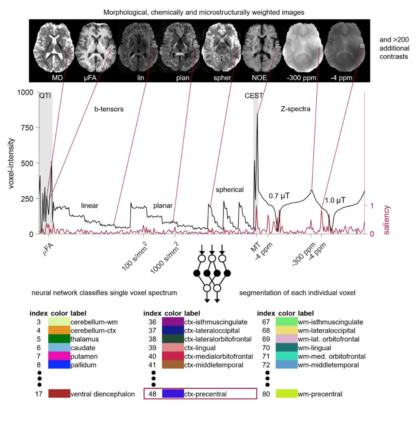
Fig. 1. Visualization of the
classification approach.