Jian Wang1, Guohui Ruan2,3, Yingjie Mei4, Yanjun Chen1, Jialing Chen1, Yanqiu Feng2,3, and Xiaodong Zhang1
1Department of Medical Imaging, The Third Affiliated Hospital of Southern Medical University, Guangzhou, China, 2Guangdong Provincial Key Laboratory of Medical Image Processing, Southern Medical University, Guangzhou, China, 3School of Biomedical Engineering, Southern Medical University, Guangzhou, China, 4China International Center, Philips Healthcare, Guangzhou, China
1Department of Medical Imaging, The Third Affiliated Hospital of Southern Medical University, Guangzhou, China, 2Guangdong Provincial Key Laboratory of Medical Image Processing, Southern Medical University, Guangzhou, China, 3School of Biomedical Engineering, Southern Medical University, Guangzhou, China, 4China International Center, Philips Healthcare, Guangzhou, China
A
fully automatic U-net model was able to segment lumbosacral plexus accurately
and shorten the segmentation time significantly.
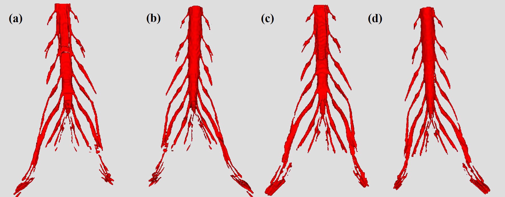
Figure
3. Segmentation results of the 3D lumbosacral
plexus. 3D model of manual segmentation (a,b) and U-Net algorithm segmentation (c,d)
of a 53 years old woman with degeneration of lumbar vertebrae and disc
herniation. The DCE, PPV and SEN between the U-net and rater is 0.867, 0.884,
and 0.850, respectively.
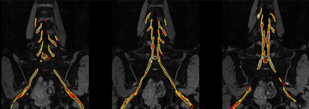
Figure
2. Segmentation results of
the 2D lumbosacral plexus. Masks of manual segmentation (red line) and U-Net
algorithm segmentation (yellow line) of a 63 years old woman with degeneration
of lumbar vertebrae. The DCE, PPV and SEN between the
U-net and rater is 0.873, 0.907, and 0.841, respectively.