Rupsa Bhattacharjee1,2, Rakesh Kumar Gupta3, Suhail P Parvaze4, Rana Patir5, Sandeep Vaishya5, Sunita Ahlawat6, and Anup Singh1,7
1Center for Biomedical Engineering, Indian Institute of Technology (IIT) Delhi, New Delhi, India, 2Philips Health Systems, Philips India Limited, Gurugram, India, 3Department of Radiology, Fortis Memorial Research Institute, Gurugram, India, 4Philips Health Systems, Philips Innovation Campus, Bangalore, India, 5Department of Neurosurgery, Fortis Memorial Research Institute, Gurugram, India, 6SRL Diagnostics, Gurugram, India, 7Department of Biomedical Engineering, All India Institute of Medical Sciences, New Delhi, India
1Center for Biomedical Engineering, Indian Institute of Technology (IIT) Delhi, New Delhi, India, 2Philips Health Systems, Philips India Limited, Gurugram, India, 3Department of Radiology, Fortis Memorial Research Institute, Gurugram, India, 4Philips Health Systems, Philips Innovation Campus, Bangalore, India, 5Department of Neurosurgery, Fortis Memorial Research Institute, Gurugram, India, 6SRL Diagnostics, Gurugram, India, 7Department of Biomedical Engineering, All India Institute of Medical Sciences, New Delhi, India
One of the first retrospective studies exploring radiomic feature
extraction and machine-learning to know whether radiomic features can
significantly differentiate between
3Dvasculature and 3DHemorrhage mask regions in
SWI-magnitude images.
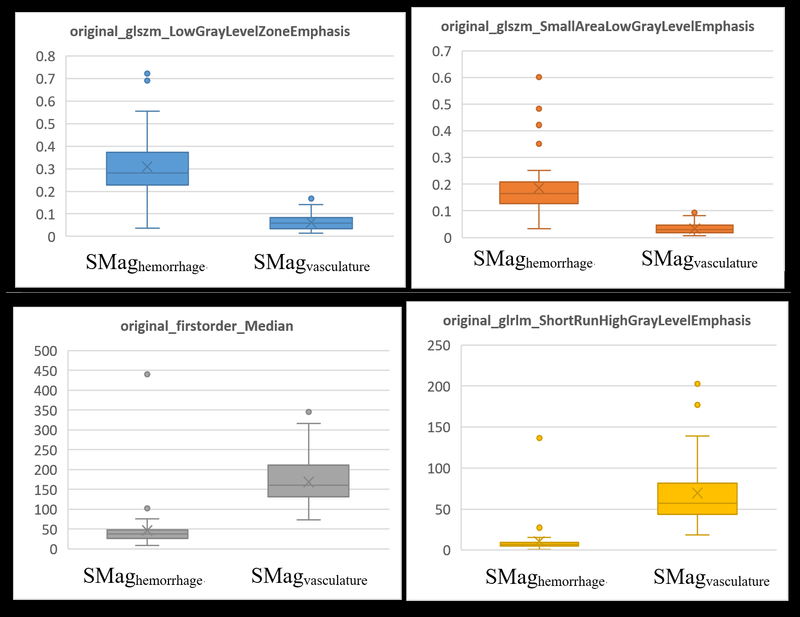
Figure-2: Representative
feature value comparisons between SMagvasculature and SMaghemorrhage
for the top-ranked four features
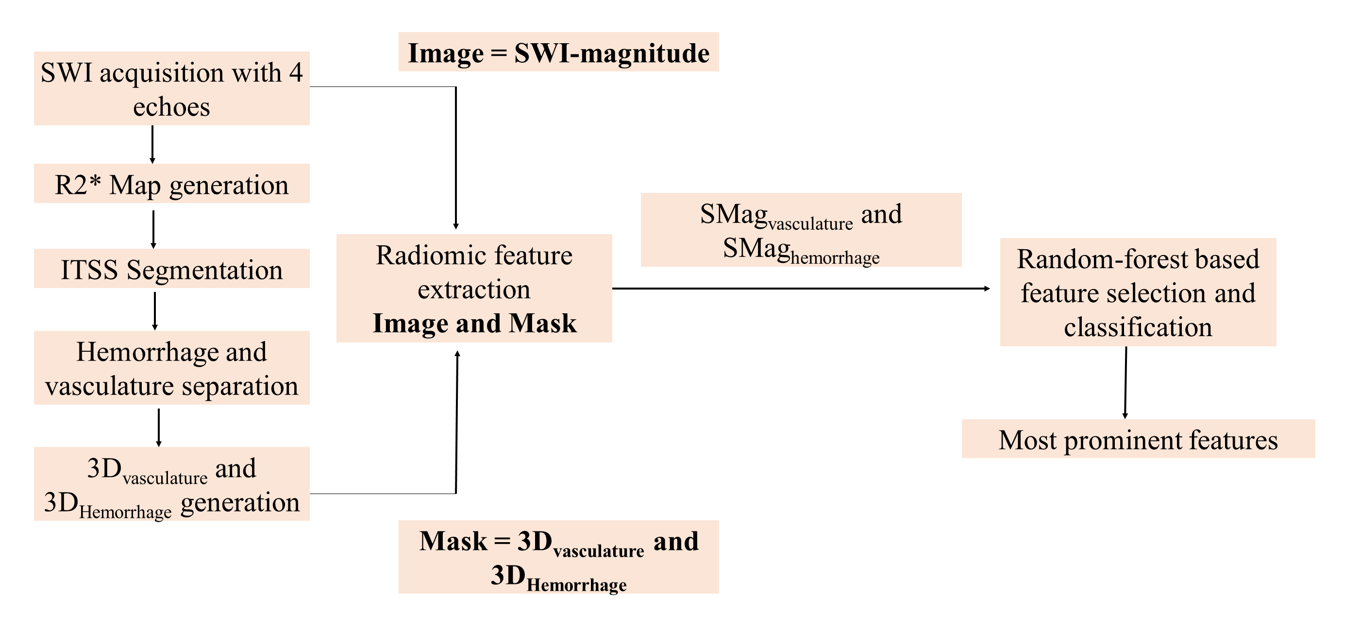
Figure-1: Flowchart
of methodology